
Page 156 - 2020_08-Haematologica-web
P. 156
L.M. Jonart et al.
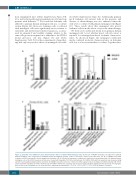
from transplanted mice (Online Supplementary Figure S1B, C) as well as histopathological examinations of brains from patients with leukemia.11.-14 We found that leukemia cells adhered to primary human meningeal cells in a co-culture system (Figure 1A). Moreover, leukemia cells co-cultured with meningeal cells were significantly more resistant to cytarabine and methotrexate-induced apoptosis, as meas- ured by annexin-V and viability staining, relative to the same cells grown in suspension or adherent to the HCN-2 neural precursor cell line (Figure 1B and Online Supplementary Table S1). In these experiments, chemother- apy had only very modest effects on meningeal cell viabil-
AB
ity (Online Supplementary Figure S2). Additionally, primary pre-B leukemia cell survival both in the presence and absence of chemotherapy was also enhanced when the cells were co-cultured with primary meningeal cells (Figure 1C). These results show that meningeal cells protect leukemia cells from the effects of cytotoxic chemotherapy.
We then used conditioned media from primary human meningeal cells to test whether direct cell-cell contact is required for meningeal-mediated leukemia chemoresis- tance. As shown in Figure 1D, meningeal conditioned media conferred moderate chemoresistance on leukemia cells, but to a lesser extent than co-culture. Together these
CD
Figure 1. Leukemia cells exhibit increased chemoresistance when co-cultured with meningeal cells. (A) Percent of NALM-6 leukemia cells adherent to primary human meningeal cells, retronectin (recombinant fibronectin fragment) positive control, or non-tissue culture treated well after 2 h. (B, C) NALM-6 and Jurkat leukemia cells (B) or primary B-cell acute lymphocytic leukemia cells (C) cultured in suspension or adherent to central nervous system-derived cells (primary human meningeal cells or the HCN-2 neuronal cell line) were treated with cytarabine 500 nM or methotrexate 500 nM for 48 h and then apoptosis was measured using annexin-V and viability staining and flow cytometry. (D) NALM-6 and Jurkat cells were cultured in either regular media or meningeal conditioned media (CM; 100%) and treated with cytarabine 500 nM or methotrexate 500 nM for 48 h before apoptosis was measured using annexin-V staining and flow cytometry. For all graphs, data are the mean ± standard error of mean from three independent experiments. **P<0.01, ***P<0.001, ****P<0.0001 by analysis of variance.
2132
haematologica | 2020; 105(8)
