
Page 161 - 2020_08-Haematologica-web
P. 161
Attenuating leukemia chemoresistance in the CNS
meninges on leukemia biology. We found that the meninges enhance leukemia resistance to cytarabine and methotrexate, the primary drugs currently used in the treatment of CNS leukemia, by altering the apoptotic bal- ance in leukemia cells to favor survival and increasing leukemia quiescence.1,2 Quiescence allows cancer cells to escape cytotoxic chemotherapy and has been shown to be critical for leukemia relapse and stem cell biology.32,51,52 In agreement, it was previously shown that high Mer kinase- expressing, t(1;19) leukemia cells co-cultured with CNS- derived cells exhibit G0/G1 cell cycle arrest, suggestive of dormancy or quiescence, as well as methotrexate resist- ance.16
To define the mechanism by which the meninges exert these effects on leukemia biology, we also tested the abil- ity of meningeal conditioned media to enhance leukemia chemoresistance. While meningeal conditioned media partially attenuated the sensitivity of leukemia cells to chemotherapy, the effect was significantly less than when leukemia cells were in direct contact with meningeal cells. This result supports a model in which leukemia chemore- sistance is primarily dependent upon direct interactions between the leukemia and meningeal cells with smaller contributions from a soluble factor(s) secreted by the meningeal cells. Together these results further support that the pathophysiology of CNS leukemia and relapse is more complex than simply the ability of leukemia cells or chemotherapy to access the restricted CNS microenviron- ment and complement other extensive laboratory and clinical data demonstrating that cell-autonomous factors play an essential role in leukemia biology.50,53,54
Importantly, we also found that meningeal-mediated leukemia chemoresistance was a reversible phenotype. Leukemia cells removed from co-culture with meningeal cells or the meninges of xenotransplanted mice reverted back to baseline cell cycle, quiescence, apoptosis balance, and sensitivity to methotrexate and cytarabine. Ebinger et al. recently identified a similar population of relapse- inducing ALL cells within the bone marrow that exhibited dormancy, stemness, and treatment resistance.31 However, similar to our results, these therapeutically adverse prop- erties were reversed when these leukemia cells were dis- sociated from the bone marrow microenvironment.
We then identified drugs capable of disrupting leukemia-meningeal adhesion. In addition to identifying several drugs that inhibit canonical cell adhesion targets, we also found that Me6TREN, a novel hematopoietic stem cell mobilizing compound, also disrupted leukemia- meningeal adhesion in vitro and in vivo. Moreover, Me6TREN enhanced the efficacy of cytarabine in treating CNS leukemia in xenotransplanted mice. In vivo efficacy against two leukemia cell lines with distinct immunophe- notypes (T- and B-cell) and a primary B-ALL patient- derived xenotransplant support the possibility that drugs, or biologic agents, that target leukemia-niche interactions may exhibit broader specificity than mutation-specific therapeutics that are limited by the genetic heterogeneity of leukemia.
The mechanism by which Me6TREN disrupts the leukemia-meningeal niche is an active area of investiga- tion in our laboratory. The ability of MMP inhibitors to diminish the efficacy of Me6TREN agrees with other work showing that Me6TREN upregulates MMP-9 in the bone marrow niche.38 Our gene expression data also showed that Me6TREN significantly downregulated the
AB
CD
E
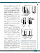
Figure 5. The effects of the meninges on leukemia cells are reversible. (A) NALM-6 and Jurkat cells grown in suspension, co-cultured with meningeal cells, or isolated from co-culture and placed back in suspension were treated with Ara- C (cytarabine) 500 nM and apoptosis was assessed by annexin-V staining and flow cytometry. (B) NALM-6 leukemia cells were isolated from the meninges of xenotransplanted mice and then grown in suspension for 48 h prior to treatment with cytarabine 500 nM and assessment of viability using annexin-V staining and flow cytometry. (C, D) NALM-6 (C) and Jurkat (D) leukemia cells were co-cul- tured with meningeal cells or isolated from co-culture and placed back in sus- pension and then cell cycle assessed using Click-iT Plus EdU cell cycle reagent and flow cytometry. (E) BH3 profiling was performed using the BIM peptide on NALM-6 and Jurkat leukemia cells co-cultured with meningeal cells or placed back into suspension after co-culture with meningeal cells. Cytochrome c reten- tion was measured by flow cytometry. For all graphs, data are the mean ± stan- dard error of mean from three independent experiments. ****P<0.0001 by analysis of variance. ns: not significant.
haematologica | 2020; 105(8)
2137
