
Page 206 - Haematologica - Vol. 105 n. 6 - June 2020
P. 206
S. Stivala et al.
AE
B
F
C
G
D
H
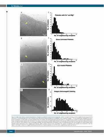
Figure 3. High-shear induces a local rearrangement of GpIb as revealed by cryo-electron tomography. (A-C) Immunogold labeling and (E-G) neighborhood analysis for the control (A, E), shear-activated (B, F) and α-linolenic acid (ALA)-treated shear-activated (C, G) platelets. (A-C): Projection images (~70 nm in thickness) obtained from tomo- grams of platelets on which GpIbα has been labeled with immunogold. The labeling was detected with protein G conjugated to 6 nm gold. To make the gold identification easier, the gold has been labeled with red circles. The yellow arrows indicate crowded neighborhoods in the pictures. All the projections have the same scale and the scale bar in A is 100 nm. (E-G) Analysis of the immunogold labeling. The three-dimensional coordinates of the 6 nm gold particles from six tomograms for each condition were selected and the distances to the “neighboring receptors” within a radius of 50 nm were calculated. The number of neighbors each receptor has is depicted in the his- togram. (D) Integrin αIIbβ3 immunogold labeling was performed on spread platelets and (H) the same neighboring analysis performed.
1664
haematologica | 2020; 105(6)
