Page 238 - Haematologica May 2020
P. 238
P.J. Teoh et al.
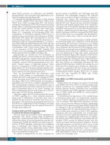
   rapid STAT3 activation (in U266-shCtr and OCIMY5- pLenti6-ADAR1) was associated with significantly accel- erated IL6-induced growth (Figure 4B).
To elucidate the potential mechanism, we first checked for a possible ADAR1-STAT3 interaction. Pulling down STAT3 from STAT3-overexpressing 293T cells showed enrichment of endogenous P150 and its known interacting partner, JAK2, in the immunoprecipitated fraction but not in the IgG control fraction. On the other hand, P110 seemed to form only a loose interaction with STAT3 (Figure 4C). Consistently, in IL6-responsive H929 cells, accumulation of IL6-induced phospho-STAT3 led to a gradual increase of interaction with endogenous ADAR1, particularly P150 (Figure 4D), indicating that it was the main interacting isoform. To confirm this observation, we performed reciprocal immunoprecipitation. Co-transfec- tion of P150-Flag and STAT3 plasmids into 293T cells and pulling down only the P150 isoform also revealed efficient co-precipitation with STAT3 protein (Figure 4E). There was no interaction of P150 with JAK2. Similarly, P150 could be seen co-precipitating with endogenous STAT3 in the IL6-induced fraction of H929 cells (Figure 4F).
Subsequently, we checked for the localization of ADAR1 and STAT3 in human MM cell lines (OCIMY5 and H929) and patients’ samples (N291 and N292), and found that STAT3 was localized in both the nucleus and cytoplasm, whereas, P150 was predominant in the cyto- plasm. Merged images of individual proteins clearly showed co-localization of STAT3 and P150 in the cyto- plasm (Figure 4G). It is therefore plausible that this cyto- plasmic co-localization could have rendered the environ- ment conducive for their physical interaction.
Next, we investigated how this interaction could enhance STAT3 activity. A cycloheximide chase assay revealed that P150 could promote the stability of STAT3 protein. Knocking down P150 enhanced STAT3 degrada- tion (Figure 4H). At 8 h, STAT3 already showed signs of degradation and was almost completely degraded at 24 h after cycloheximide treatment in the shADAR1 cells, in contrast to the shCtr cells which required up to 16 h for a mild cycloheximide effect to be visible. Similarly, the P150-overexpressed cells also took longer (up to 24 h) to show signs of STAT3 protein degradation.
These data suggest that P150 could mediate the activity of STAT3 signaling via a direct physical interaction, which leads to stabilization of the latter, thus, more is shuttled into the nucleus to transcribe various pro-onco- genic genes. This hypothesis was validated when the loss of P150 led to compromised STAT3 binding to its target gene promoters (Figure 4I) and sub-optimal induction of its target genes (Figure 4J).
Combined IL6R and P150 suppression leads to reduced IL6-induced oncogenicity
The more rapid and sustainable STAT3 signaling in the 1q21(amp) cells (which express IL6R constitutively) (Figure 1D), coupled with the STAT3- P150 feedback reg- ulatory loop (Figure 4) led us to hypothesize that the con- comitant gain of IL6R and ADAR1 may lead to hyperac- tivation of STAT3 signaling, conferring a more malignant state to 1q21(amp) cells. Indeed, the observation of 1q21(amp) patients having significantly higher STAT3 indices (STAT3 pathway activation) further supports the basis of our hypothesis (Figure 5A).
We further investigated this by first comparing the
growth profile of 1q21(WT) and 1q21(amp) upon IL6 stimulation. Not surprisingly, exogenous IL6 conferred more active growth to all tested cell lines compared to untreated ones (Figure 5B). Importantly, however, 1q21(amp) cells demonstrated more pronounced growth than the WT cells. The growth rate of the IL6-stimulated 1q21(amp) cells already surpassed that of the unstimulat- ed ones as soon as 4 h after IL6 induction. In contrast, the 1q21(WT) cells required up to 24 h after IL6 stimulation to display a differential growth phenotype. This implies that the 1q21(amp) cells had a sensitized IL6/STAT3 path- way and that there was a possibility that the pathway- related genes within 1q21 could be driving this pheno- type.
To elucidate the potential importance of IL6R and P150, we performed single and co-depletion of IL6R and P150 and assessed the functional consequences for STAT3 activity and MM cell growth. Suppression of IL6R or P150 alone obliterated the IL6-induced STAT3 pathway, mani- fested by the weaker induction of phospho-STAT3 and its downstream factors (Figure 5C). Inhibition of this path- way was detrimental to MM cells, which experienced growth retardation (Figure 5D), impeded colony forma- tion (Figure 5E) and halted IL6-induced-cell cycle progres- sion (as compared to IL6-treated shCtr cells which pro- gressed through the S/G2 phase) (Figure 5F), suggesting that both factors are individually important for IL6- induced STAT3 activation. Notably, the combined loss of IL6R and P150 resulted in more deleterious cellular phe- notypes as compared to the loss of just one respective protein, consistent with the more prominent attenuation of IL6-induced STAT3 signaling (Figure 5C). A similar trend was also observed in H929 cells (Online Supplementary Figure S6).
IL6R, ADAR1 and STAT3 demonstrate good clinical correlation
Having shown the biological association of both IL6R and ADAR1-P150 with STAT3, we then investigated the clinical correlations by analyzing publicly available patients’ datasets. Positive correlations were consistently observed between the STAT3 signatures and the expres- sion of STAT3, IL6R and ADAR1 in various patients’ datasets (Figure 6A).
To demonstrate the importance of the combined gain of both genes within 1q21, we computed the correlation between STAT3 signature enrichment and overall sur- vival in patients with differential expression of these genes. Six out of seven datasets revealed a significantly enriched STAT3 signature in the high-ADAR1+high-IL6R as compared to the low-ADAR1+low-IL6R groups of patients (Figure 6B). We also found that lack of expres- sion of either gene resulted in a lower STAT3 signature expression and, more importantly, loss of both genes caused an even more significant suppression of the signa- ture (Figure 6C), signifying that both factors are influen- tial players in the STAT3 pathway. Importantly, this dif- ferential STAT3 activity in different groups was also closely associated with the patients’ prognosis. Patients with high expression of both genes had a worse overall survival than those with low expression of both genes (Figure 6B and Online Supplementary Figure S7) further supporting our hypothesis that the concomitant gain of IL6R and ADAR1 is critical in driving poor disease out- come in the 1q21(amp) cases.
 1398
  haematologica | 2020; 105(5)


