
Page 236 - Haematologica May 2020
P. 236
P.J. Teoh et al.
A
B
C
DE
F
H
G
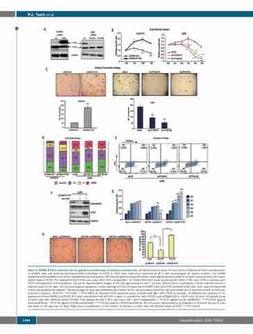
Figure 3. ADAR1-P150 is important for the growth and proliferation of multiple myeloma cells. (A) Western blot analysis to check for the efficiency of P150 overexpression in OCIMY5 cells and lentivirus-mediated-shRNA knockdown of P150 in U266 cells. Cells were harvested at 48 h after manipulation for protein isolation. The shRNA sequences are available in the Online Supplementary Information. (B) Daily cell growth assay (CTG assay) depicting the growth profile of multiple myeloma cells with manip- ulated levels of P150. The reading for day 0 (D0) was taken 48 h after manipulation. (C) Colony-formation assay assessing the ability of the cells to form colonies upon P150 overexpression and knockdown. Top panel: representative images of the soft agar incubated over 7-14 days. Bottom panel: quantification of the colonies formed in triplicate wells of soft agar. (D) Cell cycle analysis (propidium iodide staining) of P150-overexpressed OCIMY5 cells and P150-depleted U266 cells. Cells were harvested 48 h after manipulation for analysis. The percentage on each bar represents the number of the cell population within the cell cycle phase out of the total number of cells cap- tured and analyzed. *P<0.05 **P<0.001 ***P<0.0001 (E) Annexin-V-FITC apoptosis assay of U266 cells 48 h after P150 knockdown. (F) Western blot analysis for the expression of total ADAR1 and P150 24 h after transfection with shP150 or upon co-transfection with shP150 and pCDNA-P150 in U266 cells. (G) Daily cell growth assay of U266 cells with different levels of P150. The reading for day 0 (D0) was taken 48 h after manipulation. **P<0.05 against shCtr+pCDNA-EV, ***P<0.001 against shCtr+pCDNA-EV, ^^P<0.05 against shP150+pCDNA-EV, ^^^P<0.001 against shP150+pCDNA-EV. (H) Left panel: representative photographs of colonies formed in tripli- cate wells of soft agar over 14 days. Right panel: quantification of the number of colonies in U266 cells with different levels of P150. ***P<0.0001.
1396
haematologica | 2020; 105(5)
