
Page 272 - Haematologica April 2020
P. 272
L. Zheng et al.
premature stop within the propeptide, resulting in a vwf- null phenotype. Western blotting demonstrated the pres- ence of vwf multimers in plasma of wt, but not vwf-/- (Figure 6C) or a13-/-vwf-/- (not shown) zebrafish. These results demonstrate the successful deletion of the vwf gene in zebrafish.
To further assess zebrafish vwf function, we performed a microfluidic assay using whole blood from wt and vwf-/- zebrafish. When perfused through a fibrillar collagen-coat- ed surface under arterial shear, the final surface coverage of fluorescein-labeled thrombocytes (Figure 6E) and the rate of thrombocyte adhesion and aggregation (Figure 6F) onto the collagen surface were dramatically reduced in vwf-/- and a13-/-vwf-/- zebrafish compared with the wt con- trols (P<0.0001). Moreover, the time to form an occlusive thrombus, determined by intravital microscopy, in the caudal vein in vwf-/- or a13-/-vwf-/- zebrafish after FeCl3 injury was significantly prolonged when compared to that in the wt controls (Figure 6D). All wt zebrafish formed occlusive thrombi within 20 min after FeCl3 injury, but nearly 50% of vwf-/- or a13-/-vwf-/- zebrafish failed to form occlusive
thrombi. These results demonstrate the relatively defi- cient hemostatic functions in vwf-/- and a13-/-vwf-/- zebrafish.
vwf-/- and a13-/-vwf-/- zebrafish are protected
from developing spontaneous and histone-induced thrombocytopenia
Under unprovoked conditions, vwf-/- zebrafish with or without circulating plasma a13 exhibited normal counts of total (Figure 7A), immature (Figure 7B), and mature (Figure 7C) thrombocytes. Moreover, when challenged with a lysine-rich histone (H5505), vwf-/- or a13-/-vwf-/- zebrafish exhibited no significant reduction in the num- bers of total (Figure 7D, G), immature (Figure 7E, H), and mature (Figure 7F, I) thrombocytes over a 7-day period. However, 7 days after the histone challenge, there were significantly lower numbers of total (Figure 7J), immature (Figure 7K), and mature (Figure 7L) thrombocytes in a13-/- than in a13-/-vwf-/- zebrafish. Together, these results demonstrate that vwf-/- zebrafish with or without circulat- ing A13 are protected from developing spontaneous or histone-induced TTP.
ABC
DEF
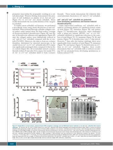
Figure 5. Kaplan-Meier survival analysis, plasma histone/DNA complexes, histology and von Willebrand factor multimers in wt and a13-/- zebrafish after histone challenge. (A) Survival rate (%) over a 2-week period in wt and a13-/- zebrafish after being challenged with phosphate-buffered saline (PBS) or lysine-rich histone (H5505) (200 mg/kg body weight). (B) Plasma levels of histone-DNA complexes in wt and a13-/- zebrafish prior to (D0) and day 7 (D7) following the histone (H5505) challenge. (C) Histological images of occlusive microvascular thrombi (arrowheads) in the liver tissue from a13-/- (top) and wt (bottom) zebrafish following lysine-his- tone (H5505) (left) or PBS challenge (right). (D) The numbers of microvascular thrombi per tissue section (box-and-whisker plot, with minimum to maximum) in wt and a13-/- zebrafish following the histone (H5505) or PBS challenge. A Mann-Whitney test was performed to test differences between two groups. *P<0.05, **P<0.01, ***P<0.005, and ****P<0.001. (E) Immunohistochemical staining showing von Willebrand factor (VWF)-rich microvascular thrombi (top, arrowheads). Scale bar represents 200 μm in length. (F) Plasma vwf multimer distribution in wt and a13-/- zebrafish prior to (D0), 1, 3, and 7 days after the histone (H5505) chal- lenge. Here, H and L on the right margin indicate high and low molecular weight VWF multimers, respectively.
1114
haematologica | 2020; 105(4)
