
Page 219 - Haematologica April 2020
P. 219
MMEJ drives 8q24 rearrangements in myeloma
Disruption of topologically associated domains by MYC rearrangements.
Topologically associated domains (TAD) have been shown to contain DNA elements that are more likely to interact with one another. Disruption of these TAD may bring super-enhancer elements into the same TAD as MYC, resulting in its increased expression. We examined the super-enhancers from the MM.1S cell line, and TAD from RPMI-8226 and U266 cell lines and integrated MYC breakpoints.
On the six frequent MYC translocation partner loci, breakpoints were clustered near to the super-enhancer and within the same TAD as the super-enhancer (Figure 6). At 8q24, the translocation breakpoints, at the two hotspots,
were clustered within the TAD containing MYC and PVT1. The resulting rearrangements would bring the super-enhancer from the partner loci adjacent to MYC, resulting in the formation of a Neo-TAD (Figure 7B) and overexpression of MYC.
We identified a patient-derived xenograft sample with a t(4;8) that resulted in insertion of three regions of chro- mosome 4 next to MYC (Figure 7A). This resulted in the super-enhancer from PCDH10, defined by the presence of H3K27Ac and MED1 marks, being placed next to MYC, resulting in overexpression. This shows for the first time in a patient sample a rearrangement that con- firms the importance of the placing of a super-enhancer next to MYC.
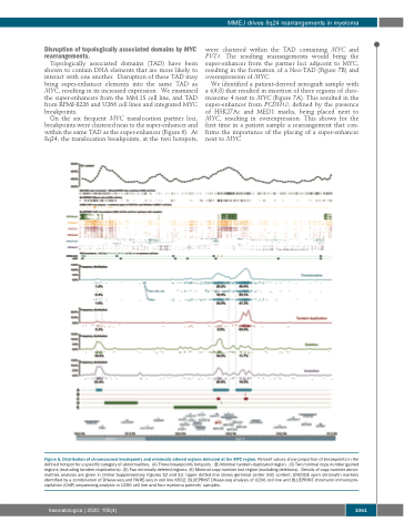
Figure 5. Distribution of chromosomal breakpoints and minimally altered regions detected at the MYC region. Percent values show proportion of breakpoints in the defined hotspot for a specific category of abnormalities. (A) Three breakpoints hotspots. (B) Minimal tandem-duplicated region. (C) Two minimal copy number gained regions (excluding tandem-duplications). (D) Two minimally deleted regions. (E) Minimal copy-number lost region (excluding deletions). Details of copy-number abnor- malities analysis are given in Online Supplementary Figures S2 and S3. Upper dotted line shows germinal center (GC) content, ENCODE open chromatin markers identified by a combination of DNase-seq and FAIRE-seq in cell line K562, BLUEPRINT DNase-seq analysis of U266 cell line and BLUEPRINT chromatin immunopre- cipitation (ChIP)-sequencing analysis in U266 cell line and four myeloma patients’ samples.
haematologica | 2020; 105(4)
1061
