
Page 217 - Haematologica April 2020
P. 217
MMEJ drives 8q24 rearrangements in myeloma
RNA-sequencing data, being present in >95% of patients with log2 normalized counts >10. All of the loci except for IGK had super-enhancers previously identified in the MM.1S cell line; 67.2% (205 of 305) of cases with non- complex translocation (5 or less loci involved) had at least one of these super-enhancers involved in the transloca- tion. Another five partners were present in 5-10 cases, three of which overlapped with the highly-expressed genes FCHSD2, FBXW7 and SERTAD2, which are associ- ated with known super-enhancers.30
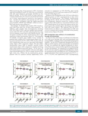
Interestingly, 13 samples had complex MYC transloca- tions with more than one of these super-enhancers. In addition, eight samples had rearrangements involving IGH, MYC and CCND1, and four samples had rearrange- ments with IGH, MYC and MAF, indicating that they may occur as primary events early in the disease process. All oncogenes involved in these translocations show high expression (Online Supplementary Figure S5). This targeting of multiple oncogenes may explain worse survival in patients with complex MYC translocations (Figure 1B). Ig loci were involved in 47.9% (146 of 305) of cases with an MYC translocation and were not associated with signifi- cantly higher MYC expression (Figure 3B and Online Supplementary Figure S6B) or patients’ survival (Online Supplementary Figure S4D) compared to samples involving other super-enhancer-associated genes. In six cases, an IGH translocation occurred together with one of the light- chain immunoglobulin loci, but no sample involved both light chain loci. Within the Ig translocation groups, patients with IGL partners showed significantly worse
outcome in comparison to IGH (P<0.05), other non-Ig translocations (P<0.01), and cases without MYC transloca- tions (P<0.001) (Figure 1C).
Analysis of the breakpoints at the IGH locus indicated a different pattern of MYC rearrangements to that of the primary Ig translocations. The primary translocations involving t(4;14), t(6;14), t(11;14), t(14;16), and t(14;20) have breakpoints clustered around the constant switch regions where AID motifs are concentrated. However, the MYC translocations do not share this pattern and are dis- persed across the constant region, showing no association with AID motif clusters. This indicates that the MYC translocations are likely to be independent of AID and occur in a manner that is distinct from that of the primary translocations (Figure 4A and B).
MYC breakpoints show evidence of recombination through microhomology
It is known that class switch recombination breakpoints in B cells occur through AID and non-homologous end joining (NHEJ), resulting in blunt ended DNA being ligated together.33 As the MYC breakpoints identified here do not align to switch regions, and are presumably not mediated by AID, we examined the aligned breakpoints to deter- mine if they were constructed through blunt ended joining or other mechanisms. In comparison to re-aligned t(4;14), t(6;14), t(11;14), t(14;16), and t(14;20) breakpoints, which are mediated by AID and NHEJ, the MYC breakpoints had significantly fewer blunt ended rearrangements (54.1% vs. 27.7%; P<0.001) and significantly more rearrangements
ABC
DEF
Figure 3. RNA-sequencing expression analysis of MYC and PVT1 in relation to chromosomal abnormalities at 8q24. Effect of abnormality type (A and D), translo- cation category (B and E), and translocation breakpoint position (C and F) are shown for MYC and PVT1, respectively. *P<0.05; **P<0.01; ***P<0.001. n: number.
haematologica | 2020; 105(4)
1059
