
Page 53 - 2019_12-Haematologica-web
P. 53
Disruption of MBD2-NuRD induces high HbF levels
expression of WT or mutant MBD2 in MBD2KO HUDEP- 2 cells. For these studies, three lentiviral MBD2 expres- sion vectors were engineered. ‘MBD2sgR’ is a WT MBD2 construct with translationally silent mutations in the GR domain designed to convey resistance to cleavage by Cas9 (denoted as the sgR mutation). The ‘CCmutsgR’ construct contains the sgR mutation along with three amino acid substitutions (D366R/R375E/R380E) in the CC domain of MBD2. ‘IDRmutsgR’ contains both the sgR mutation and two sequential amino acid substitutions (R286E/L287A) in the IDR of MBD2 (Figure 4A and Online Supplementary Figures S7-S10). MBD2KO HUDEP-2 cells were infected with each of these constructs. The level of exogenously expressed MBD2 proteins was closely matched with endogenous MBD2 expression seen in the scrambled guide (sgSCR + empty vector) control cells (Figure 4C). When WT MBD2 (MBD2sgR) was added back to the MBD2KO HUDEP-2 cells, there was a 5-fold reduction in relative g-globin expression and a decrease in g/g+β mRNA level compared to the MBD2KO + empty vector control (Figure 4D and E). This corresponds to a significant rescue of the WT MBD2 phenotype compared to the empty vec- tor control. In contrast, neither the CC or IDR mutant restore g-globin gene silencing. In order to determine whether the CC mutation and IDR mutation selectively cause dissociation of the CHD4 and HDCC subcompo- nents, or disrupt the entire complex, we tested the ability of these mutants to pull down NuRD components by per- forming co-immunoprecipitation in 293T cells. The CCmutsgR construct pulled down almost no GATAD2A or CHD4, but did pull down similar amounts of MTA2 and HDAC2 compared to the MBD2sgR construct. Conversely, the IDRmutsgR construct pulled down less MTA2 and HDAC2 compared to the MBD2sgR construct, but did pull down similar levels of CHD4 and GATAD2A (Figure 4B), consistent with our structural predictions. Together these data provide strong evidence that pertur- bation of either of these two interaction domains is suffi- cient to functionally diminish MBD2-NuRD mediated g-globin gene silencing by independently decoupling one of the NuRD subcomplexes.
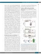
The R286E/L287 mutation of the MBD2 intrinsically disordered region disrupts helical propensity
two helical regions, showing that the R286E/L287A muta- tion disrupts the structural propensity of the IDR, thereby reducing its ability to bind the HDCC (Figure 5C).
Based on previous structural analyses,27 the CC muta- tions involve residues that form critical interactions with GATAD2A. However, the structure of the IDR bound to the HDCC has yet to be determined, and it is unclear whether the IDR mutations involve residues that make direct contact with HDCC components or if they reduce the structural propensity of the IDR and thereby indirectly disrupt binding. We previously demonstrated, based on NMR chemical shift analyses, that the IDR contains three regions with inherent helical propensity.25 The double mutation occurs within the first of these regions, as high- lighted in Figure 5B. To test whether this mutation dis- rupts the helical propensity of the IDR, we assigned the NMR resonances for the mutant domain and compared 13Ca and 13C’ chemical shifts with the WT IDR. The 15N-HSQC spectrum of the mutant IDR is nearly identical to that of the WT (Figure 5A). Differences in 13Ca and 13C’ chemical shifts between WT and mutant IDR (effec- tively the difference in chemical shift index) are plotted in Figure 5B. This analysis reveals a positive deviation in chemical shifts (wild-type – mutant) throughout the first
Figure 5. The R286E/L287A double mutation reduces the helical propensity of the intrinsically disordered region (IDR). (A) Wild-type (WT) and mutant spectra. An overlay of the 2D 15N-HSQC spectra is shown for the WT (red) and
Knockdown of MBD2 in primary human CD34+ erythroid progenitor cells strongly up-regulates g-globin expression across different levels of erythroid differentiation
Comparison of the relative effects of depleting g-globin gene silencers in primary CD34+ progenitor-derived ery- throblasts on g/g+β globin mRNA levels is confounded by assay conditions at inconsistent stages of differentia- tion.9,13-15,27 To address this, we carried out shRNA MBD2 knockdown in primary CD34+ progenitor-derived ery- throblasts, and harvested mRNA at day 5 and day 7 of ery- throid differentiation. This resulted in a consistent approx- imately 10-fold increase in g/g+β mRNA, and a level of 40% g/g+β compared to 4% in scramble controls after five days of differentiation (Figure 6A and B).
A
B
C
R286E/L287A (blue) MBD2-IDR. (B)
C chemical shift changes. Bar graphs
13
(C ) between WT and R286E/L287A MBD2-IDR (WT – mutant in ppm). Three
depict the differences in chemical shift of the carbonyl (C’) and for a-carbons
regions that show helical propensity are indicated with blue ellipses and the site of mutation indicated with red squares. Positive chemical shift changes indicate that the mutant MBD2-IDR shows less helical propensity in the region surround- ing the site of mutation. (C) The results suggest that the R286E/L287A muta- tion disrupts binding to the histone deacetylase core of NuRD by reducing inher- ent helical propensity.
a
haematologica | 2019; 104(12)
2367
