
Page 54 - 2019_12-Haematologica-web
P. 54
X. Yu et al.
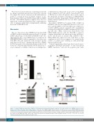
We observed a robust induction of g-globin protein with no change in β-globin protein level assayed by western blot (Figure 6C), and no aberration in the differentiation profile, as assayed by flow cytometric analysis of ery- throid markers CD71 and CD235a (Figure 6D), in MBD2 Kd cells. Thus MBD2 depletion (approx. 80%) does not impede the overall developmental stage or differentiation state of primary erythroblasts, independently validating the results in HUDEP-2 cells.
Discussion
Here we demonstrate that CRISPR/Cas9 mediated KO of MBD2 results in markedly increased levels of g-globin mRNA and protein as well as HbF in HUDEP-2 cells. The approximately 50% g/g+β mRNA level of g induction in MBD2KO HUDEP-2 cells is comparable to the effect seen with KO of BCL11A or LRF, two of the strongest g-globin gene silencers reported.13 Similarly knockdown of MBD2 in CD34+ progenitor-derived primary human erythroid cell results in a consistent approximately 10-fold increase in % g/g+β compared to scramble controls across multiple days
of differentiation. This results in up to 40% g/g+β mRNA levels, compared to 4-5% in controls. Given the ≥5% level of HbF in most sickle cell patients, these results support the therapeutic potential of disruption of MBD2-NuRD- mediated silencing. Importantly, MBD2 knockdown in primary human erythroid cells does not affect erythroid differentiation.
In contrast, CRISPR/Cas9 mediated KO of MBD3 in HUDEP-2 cells does not appreciably increase g/g+β or rel- ative g-globin mRNA or protein expression compared to scramble sgRNA controls, consistent with our published observation of siRNA Kd of MBD3 in human β-YAC bear- ing CID cells.27 Other studies have demonstrated that MBD3-NuRD interacts with the TR2/TR4 co-repressor complex which binds the embryonic β-type globin pro- moter39 and BCL11A in murine MEL erythroid cells.40 While MBD3 may indeed associate with these or other complexes in vivo, it does not appear to be essential in the context of g-globin gene silencing in adult phenotype human erythroid cells.
Genomic engineering technologies have been shown to recapitulate hereditary persistence of fetal hemoglobin (HPFH) mutations,38 and edit the β-globin gene sickle
AB
CD
Figure 6. Lentiviral shRNA knockdown (Kd) of MBD2 in CD34 progenitor-derived primary human erythroid cells results in high level g/g+β RNA expression and g- globin protein without affecting erythroid differentiation. (A) Relative Kd of MBD2 mRNA’. (B) approximately 10-fold increase in g/g+β mRNA in MBD2 kd primary erythroid cells, across two different levels of differentiation, compared to scrambled shRNA controls. (C) Increase in g-globin protein without change in β-globin protein as measured by western blot using anti γ-globin and anti β-globin antibody. (D) Flow cytometry analysis showing equivalent erythroid differentiation profiles of scram- ble control (sc) and MBD2 Kd CD34+ cells at day 7. Error bars represent ± Standard Deviation of three biological repeats. *P<0.05; **P< 0.01; n.s.: P>0.05. Statistical testing was performed using the Student’s t-test.
2368
haematologica | 2019; 104(12)
