
Page 52 - 2019_12-Haematologica-web
P. 52
X. Yu et al.
globin mRNA compared to scrambled guide RNA control cells (Figure 3B and C) and no difference in relative g-glo- bin mRNA (Online Supplementary Figure S6A). MBD3KO clone9.5 showed a minimal but statistically significant increase to 0.31% g/g+β compared to 0.13% in the sgSCR population with a correlative increase in relative g-globin mRNA level (Figure 3C and Online Supplementary Figure S6A); however, these effects were very small and inconsistent compared to those for MBD2KO cells. Importantly, we observed no detectable increase in g-glo- bin protein by western blot compared to scramble for any MBD3KO clone, while the MBD2KO pooled line showed robust g-globin protein expression (Online Supplementary Figure S1B). Relative β-globin mRNA levels trended slight- ly higher in MBD3KO cells compared to scramble controls (Online Supplementary Figure S6B); this was reflected at the protein level (Online Supplementary Figure S1B). These results provide strong evidence that MBD3-NuRD is not an important mediator of g-globin gene silencing in human erythroid cells.
Amino acid substitutions in the intrinsically disordered region and C terminal coiled-coil domains of MBD2 that disrupt MBD2-NuRD component interactions prevent g-globin gene repression in HUDEP-2 cells
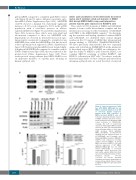
The coiled-coil (CC) domains of MBD2 and GATAD2A interact to form a stable heterodimeric complex, and this interaction is necessary for the recruitment of GATAD2A and CHD4 to the MBD2-NuRD complex.27 By determin- ing the structure and binding dynamics between MBD2 and GATAD2A, we identified three critical charged residues in the CC region of MBD2 that when mutated disrupt binding to GATAD2A (D366R/R375E/R380E).41 We have previously demonstrated that two sequential amino acid substitutions (R286E/L287A) in the intrinsical- ly disordered region (IDR) of MBD2 are sufficient to dis- rupt the ability of MBD2 to pull down the HDAC core complex (HDCC) consisting of MTA2, RBBP4/7, and HDAC2, collectively in 293T cells.25 In order to study the functional importance of these domains and interactions in human erythroid cells, we tested the effect of enforced
A
BC
DE
Figure 4. Enforced expression of wild- type (WT) MBD2 (MBD2sgR) but not MBD2 containing mutations in its IDR or coiled-coil domain suppresses gamma globin RNA expression in MBD2 knockout HUDEP-2 cells. (A) Schematic depicting domains mutated in MBD2 lentiviral expression vectors. Silent mutations in the GR domain (sgR) convey resistance to CRISPR/Cas9 cleavage. (B) Co-IP of exogenously expressed FLAG-tagged MBD2 mutant contructs in 293T cells using anti-FLAG shows differing abilities to pull down NuRD members CHD4, GATAD2A, MTA2, and HDAC2. (C) Western blot showing enforced expression levels of WT MBD2sgR, and CCmutsgR, and IDRmutsgR MBD2 mutants in MBD2KO HUDEP-2 cells compared to scramble control cell levels of MBD2. (D and E) MBD2 knockout HUDEP-2 cells with enforced expression of WT MBD2 (MBD2KO+MBD2sgR) but not IDR- mutant (IDRmutsgR) or CC-mutant (CCmutsgR), causes decreased g/g+β and relative g-globin mRNA. Error bars represent ± Standard Deviation of three biological repeats. *P<0.05; **P<0.01; n.s.: P>0.05. Statistical testing was performed using analysis of variance followed by the Tukey’s honest- ly significant difference procedure post- hoc test.
2366
haematologica | 2019; 104(12)
