
Page 208 - 2019_12-Haematologica-web
P. 208
M. Benagiano et al.
AB
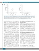
Figure 3. Cytokine profile of atherosclerotic plaque β2GPI-specific CD4+ T-cell clones obtained from systemic lupus erythematosus patients positive for antiphos- pholipid antibodies. Th clones were tested for cytokine production (A and B). β2GPI-specific Th clones were stimulated with β2GPI and TNF-a and IL-4, IFN-g and IL-17 production was measured in culture supernatants. In unstimulated cultures, levels of TNF-a, IL-4, IFN-g and IL-17 were consistently < 20 pg/mL. CD4+ T-cell clones producing IFN-g, but not IL-17 nor IL-4 were coded as Th1. CD4+ T-cell clones producing IL-17, but not IFN-g nor IL-4 were coded as Th17. CD4+ T-cell clones producing IFN-g, and IL-17, but not IL-4 were coded as Th17/Th1. CD4+ T-cell clones producing TNF-a and IL-4, but not IL-17 were coded as Th0.
β2GPI-specific atherosclerotic lesion-infiltrating T cells help monocyte tissue factor production and procoagulant activity
Plaque rupture and consequent thrombosis are crucial complications of atherosclerosis. TF plays a key role in triggering atherothrombotic events being the primary acti- vator of the coagulation cascade. We investigated whether atherosclerotic lesion-infiltrating β2GPI-specific T cells had the potential to express helper functions for TF pro- duction and PCA by autologous monocytes. Antigen- stimulated β2GPI-specific atherosclerotic lesion-derived T-cell clones were co-cultured with autologous monocytes and levels of TF and PCA were measured. Antigen stimu- lation resulted in the expression of substantial help for TF (Figure 5A) production and PCA (Figure 5B) by autologous monocytes.
Atherosclerotic lesion-derived β2GPI-specific T-cell clones express antigen-dependent help to autologous B cells for Ig production
T/B-cell interaction is a multistep process resulting in B-cell help depending on the functional commitment of the Th cells involved. So far the ability of SLE-APS-derived β2GPI-specific T-cell clones to provide B-cell help for Ig synthesis has been investigated. In the absence of the spe- cific antigen, no increase in IgM, IgG, or IgA production above spontaneous levels measured in cultures containing B cells alone was observed. In the presence of β2GPI and at a T-to-B cell ratio of 0.2 to 1, all of the β2GPI-specific T-cell clones provided substantial help for Ig production. At a 1:1 T/B cell ratio, β2GPI-dependent T-cell help for IgM, IgG, and IgA production by B cells was much higher (Figure 6). However, at a 5:1 T/B cell ratio, co-culturing B cells with autologous β2GPI-specific T-cell clones in the presence of β2GPI resulted in a much lower Ig synthesis.
Atherosclerotic lesion-derived β2GPI-specific T-cell clones display cytotoxic and pro-apoptotic activity
The cytolytic potential of SLE-APS-derived atheroscle- rotic lesion-derived β2GPI-specific autoreactive T-cell
derived from the atherosclerotic lesions showed prolifera- tion to β2GPI (Online Supplementary Table S2). A total number of 135 CD4+ and 21 CD8+ T-cell clones were obtained from atherosclerotic lesions of five SLE aPL-pos- itive. For each patient, CD4+ and CD8+ atherosclerotic lesion-derived T-cell clones were assayed for proliferation
++ in response to medium or β2GPI. 25 CD4 and no CD8
T-cell clones derived from the atherosclerotic lesions of SLE aPL-positive patients showed proliferation to β2GPI (Online Supplementary Table S3). A total number of 136 CD4+ and 30 CD8+ T-cell clones were obtained from ath- erosclerotic lesions of five SLE aPL-negative. For each patient, CD4+ and CD8+ atherosclerotic lesion-derived T- cell clones were assayed for proliferation in response to medium or β2GPI. None of the CD4+ or CD8+ T-cell clones derived from the atherosclerotic lesions showed proliferation to β2GPI (Online Supplementary Table S4).
All β2GPI-specific T-cell clones, both those obtained from the atherosclerotic lesions of SLE-APS patients and those obtained from SLE aPL-positive patients, were stim- ulated with β2GPI and autologous APC. Then, TNF-a and IL-4, IFN-g and IL-17 production was measured in culture supernatants. Upon antigen stimulation with β2GPI of the 71 β2GPI-specific T-cell clones obtained from SLE-APS patients, 30 were polarized Th1 clones, 10 Th clones were Th17, 27 Th clones were Th17/Th1, and only 4 were able to produce IL-4 together with TNF-a (Th0 clones) (Figure 2). Upon antigen stimulation with β2GPI of the 25 β2GPI- specific T-cell clones obtained from SLE aPL-positive patients, 10 were polarized Th1 clones, 6 Th clones were polarized Th17, 8 Th clones were Th17/Th1, and only one was Th0 (Figure 3). T-cell blasts from each of the 71 β2GPI-reactive T-cell clones obtained from atherosclerotic lesions of patients with SLE-APS were further screened by IFN-g and IL-17 ELISPOT in response to β2GPI. Upon appropriate stimulation, 61 atherosclerotic-derived CD4+ T-cell clones produced IFN-g, and thirty-seven produced IL-17 (Figure 4). Interestingly, all IL-17-producing β2GPI- reactive T-cell clones, produce IL-21 (mean±SE, 3.3±0.5 ng/mL per 106 T cells) in response to antigen stimulation.
2522
haematologica | 2019; 104(12)
