
Page 210 - 2019_12-Haematologica-web
P. 210
M. Benagiano et al.
and pro-coagulant β2GPI-specific Th17, Th1 and Th17/Th1 infiltrate in human atherosclerotic lesions of patients with SLE-APS and may represent a key pathogen- ic atherothrombotic mechanism.
Many self antigens, such as oxLDL, may theoretically be involved in SLE-APS atherosclerosis; oxLDL-specific peripheral blood-derived T cells displaying a Th1 profile were reported in APS patients.33 However, there is no information on whether these cells are actively involved in atherosclerotic tissue lesions of SLE-APS patients. In addition, β2GPI was found to bind ox-LDL34 raising the issue of whether or not the immune response is against ox-LDL or β2GPI itself.
The relevance of the data presented in this paper con- sists in the demonstration that all ten SLE-APS patients with clinically severe atherothrombosis harbored in their target tissues, such as atherosclerotic lesions, in vivo-acti- vated CD4+ T cells able to react to β2GPI. CD4+ T cells specific for β2GPI were found also in the plaques of SLE aPL-positive patients but not in SLE aPL-negative patients
nor in atherosclerotic patients without SLE. The results suggested that β2GPI drive inflammation in atherosclerot- ic plaques in SLE-APS and SLE aPL-positive patients, while in SLE aPL-negative patients and in non-SLE patients other antigens are involved in sustaining plaque inflammation. With the experimental procedure used in this study, the proportion of β2GPI-specific CD4+ T-cell clones generated from atherosclerotic plaques of atherothrombotic SLE- APS patients is remarkably higher than the frequency of β2GPI-specific T cells found in their peripheral blood.
In order to investigate plaque instability, we investigat- ed fresh T cells coming from the atherosclerotic plaques of SLE-APS patients and we found that plaque-derived CD4+ T cells specifically produce IFN-g and IL-17 in response to both β2GPI and to mitogen stimulation. Studying at clonal level the β2GPI-specific T cells found in the inflammatory atherosclerotic infiltrates of SLE-APS we found that 42% were polarized T helper 1 cells, 38% were Th17/Th1 cells, 15% were polarized Th17 cells, 5% were Th0 cells, and no T cells were polarized Th2 cells. The lack of Th2 cells
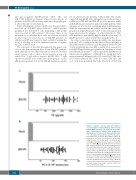
A
B
Figure 5. Induction of tissue factor (TF) synthesis and procoagulant activity (PCA) by atherosclerotic plaque β2GPI-specific T cells derived from systemic lupus erythematosus patients with antiphospho- lipid syndrome. Atherosclerotic plaque β2GPI-specif- ic T cells induce TF production and PCA by autolo- gous monocytes. To assess their ability to induce TF production and PCA by autologous monocytes, β2GPI-specific Th clones were co-cultured with autologous monocytes in the presence of medium () or β2GPI () (A). TF production by monocytes was assessed by ELISA. The results shown represent TF levels induced by T-cell clones over the TF produc- tion in cultures of monocytes alone. Atherosclerotic plaque-derived β2GPI-specific T-cell-induced PCA in autologous monocytes (B). β2GPI-specific Th clones were co-cultured with autologous monocytes in the presence of medium () or β2GPI (). At the end of the culture period, cells were disrupted and total PCA was quantitated as reported in the Methods section. The results shown represent PCA induced by T-cell clones in monocytes over the PCA in cul- tures of monocytes alone.
2524
haematologica | 2019; 104(12)
