
Page 209 - 2019_12-Haematologica-web
P. 209
Th17/Th1 inflammation in lupus atherosclerosis
A
B
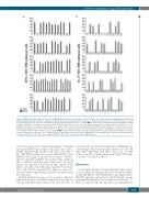
Figure 4. β2GPI driven IFN-g and IL-17 secretion by β2GPI-specific atherosclerotic plaque derived Th clones from systemic lupus erythematosus patients with antiphospholipid syndrome. Numbers of IFN-g spot-forming cells (SFC) after stimulation of atherosclerotic plaque derived T-cell clones with medium alone, or β2GPI (A). T-cell blasts from each clone were stimulated for 48 hours (h) with medium alone (), or β2GPI (), in the presence of irradiated autologous antigen-presenting cell (APC) in ELISPOT microplates coated with anti-IFN-g antibody. IFN-g SFC were then counted by using an automated reader. After specific stimulation, 61 of 71 β2GPI-specific atherosclerotic plaque-derived T-cell clones produced IFN-g. Values are mean±Standard Deviation (SD) number of SFC per 105 cultured cells over back- ground levels. Numbers of IL-17 SFC after stimulation of atherosclerotic plaque derived T-cell clones with medium alone, or β2GPI (B). T-cell blasts from each clone were stimulated for 48 h with medium alone (), or β2GPI () in the presence of irradiated autologous antigen-presenting cells in ELISPOT microplates coated with anti-IL-17 antibody. IL-17 SFC were then counted by using an automated reader. After specific stimulation 37 of 71 β2GPI-specific atherosclerotic plaque-derived T-cell clones produced IL-17. Values are mean±SD number of SFC per 105 cultured cells over background levels.
clones was assessed by using antigen-pulsed 51Cr-labeled autologous EBV-B cells as targets. At an E:T ratio of 10:1, all Th1 and Th17/Th1 specific T-cell clones were able to lyse β2GPI-presenting autologous Epstein-Barr virus (EBV)-B cells (range of specific 51Cr release, 35-65%), whereas autologous EBV-B cells pulsed with control ag and co-cultured with the same clones were not lysed (Figure 7A). Likewise 2 Th0 and all Th17 specific T-cell clones were able to lyse their target (specific 51Cr release: 50% and 25-45% respectively), while no lysis was observed when using autologous EBV-B cells pulsed with the control ag.
Fas-FasL mediated apoptosis was assessed using Fas+ Jurkat cells as target. T-cell blasts from each clone were co- cultured with 51Cr-labeled Jurkat cells at an E:T ratio of
10,5,and2.5to1for18hinthepresenceofPMAandion- omycin (Figure 7B). Upon mitogen activation, 27 out of 30 Th1, 24/27 Th17/Th1, 4/10 Th17, and 2 out of 4 Th0 clones were able to induce apoptosis in target cells (range of specific 51Cr release: 25-61%).
Discussion
Several clinical studies and experimental models suggest a role for aPL in accelerating atherosclerotic plaque forma- tion in SLE. On the other hand, there is growing evidence that aPL represent a risk factor for arterial thrombosis sup- porting their pathogenic role in cardiovascular events.1,3,4,32 Here, we report for the first time that a pro-inflammatory
haematologica | 2019; 104(12)
2523
