
Page 207 - 2019_12-Haematologica-web
P. 207
Th17/Th1 inflammation in lupus atherosclerosis
AB
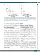
Figure 2. Cytokine profile of atherosclerotic plaque β2GPI-specific CD4+ T-cell clones obtained from systemic lupus erythematosus patients with antiphospholipid syndrome. Th clones were tested for cytokine production (A and B). β2GPI-specific Th clones were stimulated with β2GPI and TNF-a and IL-4, IFN-g and IL-17 produc- tion was measured in culture supernatants. In unstimulated cultures, levels of TNF-a, IL-4, IFN-g and IL-17 were consistently < 20 pg/mL. CD4+ T-cell clones producing IFN-g, but not IL-17 nor IL-4 were coded as Th1. CD4+ T-cell clones producing IL-17, but not IFN-g nor IL-4 were coded as Th17. CD4+ T-cell clones producing IFN-g, and IL-17, but not IL-4 were coded as Th17/Th1. CD4+ T-cell clones producing TNF-a and IL-4, but not IL-17 were coded as Th0.
APS, although they were positive for aPL, with serum anti-β2GPI, anti-cardiolipin antibodies or with positivity for LA. All SLE aPL- neg patients were affected by SLE but not by APS, and they were triple negative for serum aPL, such as anti-β2GPI, anti-cardiolipin antibodies and with negativity for Lupus Anticoagulant.
Generation and characterization of T-cell clones from atherosclerotic plaques inflammatory infiltrates
Carotid specimens, obtained by endoarterectomy, were investi- gated in both SLE-APS and in aPL negative patients under the same experimental conditions. Specimens were then disrupted, and single T cells were cloned under limiting dilution, as described.16 To assess their phenotype profile, T-cell clones were screened by flow cytometry with fluorochrome-conjugated anti- CD3, anti-CD4, anti-CD8 on a BD FACSCanto II (BD Bioscience), using the FACS Diva 6.1.3. software. The repertoire of the TCR Vβ chain of β2GPI-specific Th clones was analyzed with a panel of mAb specific to the following: Vβ1, Vβ2, Vβ4, Vβ5.1, Vβ5.2, Vβ5.3, Vβ7, Vβ8, Vβ9, Vβ11, Vβ12, Vβ13.1, Vβ13.2 and Vβ13.6, Vβ14, Vβ16, Vβ17, Vβ18, Vβ20, Vβ21.3, Vβ22, and Vβ23 (Beckman Coulter); Vβ6.7 (Gentaur) and Vβ3.1 (In Vitro Gen). Isotype-matched non-specific Ig were used as negative control. Vβ10, Vβ15, and Vβ19 T-cell receptor typing were investigated by Clontech kit, according to the manufacturer's instructions. Each β2GPI-reactive CD4+ T-cell clone was stained by only one of the TCR-Vβ chain–specific monoclonal antibodies, showing a single peak of fluorescence intensity (Online Supplementary Figure S1). The cytokine production, the cytotoxicity, the helper functions for antibody and tissue factor production of β2GPI-specific T-cell clones were performed as described.16,30, 31
Statistical analysis
Statistical analyses were performed using Student’s t-test. P<0.05 was considered significant.
Results
Atherosclerotic lesions of systemic lupus
erythematosus patients with antiphospholipid
syndrome and systemic lupus erythematosus patients
Anti-phospholipid antibody detection
The detection of aCL and aβ2GPI in patient sera, and analysis of LA was performed as described elsewhere.28,29
Atherosclerotic plaque-infiltrating in vivo activated T cells were expanded in vitro in an hrIL-2 conditioned medi- um, subsequently cloned and studied for their phenotypic and functional profile. A total number of 297 CD4+ and 37 CD8+ T-cell clones were obtained from atherosclerotic lesions of ten SLE-APS patients. For each patient, CD4+ and CD8+ atherosclerotic lesion-derived T-cell clones were assayed for proliferation in response to medium, or β2GPI. None of the CD8+ T-cell clones showed proliferation to β2GPI although they proliferated in response to mitogen stimulation (Figure 1). We have also investigated the amount of β2GPI-specific T cells present in the peripheral blood of SLE-APS patients and compared it with the one found in atheromas. The proportion of β2GPI-specific CD4+ T-cell clones generated from atherosclerotic plaques of SLE-APS patients was 24%, which is remarkably higher than the frequency of β2GPI-specific T cells found in the peripheral blood of the same patients (between 1:1900 and 1:3400).
Seventy-one (24%) of the 297 CD4+ T-cell clones gener- ated from SLE-APS atherosclerotic plaque-infiltrating T cells proliferated significantly to β2GPI (Figure 1). Each SLE-APS patient displayed a comparable percentage of CD4+ T-cell clones responsive to β2GPI (Online Supplementary Table S1). On the other hand, a total num- ber of 288 CD4+ and 42 CD8+ T-cell clones were obtained from atherosclerotic lesions of ten atherothrombotic patients, that were negative for aPL. For each patient, CD4+ and CD8+ atherosclerotic lesion-derived T-cell clones were assayed for proliferation in response to medi- um or β2GPI. None of the CD4+ or CD8+ T-cell clones
+
autoreactive β2GPI-specific CD4 T-cell clones
positive for antiphospholipid antibodies harbor
haematologica | 2019; 104(12)
2521
