
Page 206 - 2019_12-Haematologica-web
P. 206
M. Benagiano et al.
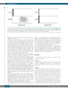
A
B
Figure 1. Antigen specificity of atherosclerotic plaque CD4+ T and CD8+ T-cell clones obtained from systemic lupus erythematosus patients with antiphospholipid syndrome. Both CD4+ T- and CD8+ T-cell clones were tested for antigen-specificity. T-cell clones were analyzed for their responsiveness to β2GPI (10 nM) (), or medi- um () by measuring [3H]thymidine uptake after 60 hours of co-culture with irradiated autologous peripheral blood mononuclear cells. Seventy-one out of 297 CD4+ T-cell clones proliferated in response to β2GPI and are shown in (A). None of the 37 CD8+ T-cell clone proliferated to β2GPI (B).
Bulkley et al.5 in a necroscopic study, that was further con- firmed by Urowitz et al.6
Many studies showed that SLE is associated with coro- nary heart disease and atherosclerosis;7-9 an important prospective study demonstrated that SLE patients have an accelerated progression of carotid plaque formations com- pared to non-lupus controls.10 SLE patients have a reduced life expectancy mainly due to the increased prevalence of cardiovascular diseases. Incidence of major cardiovascular events is 2.5 times higher in SLE patients compared to the general population. Compared to healthy subjects, SLE women, aged 35-44 years, have a 50 times increased risk of myocardial infarction and accelerated atherosclerosis, that is a well recognized comorbidity in SLE.11,12
Atherosclerosis is a multifactorial disease for which a number of different pathogenic mechanisms have been proposed. In addition to classical risk factors, in the last two decades, attention has been focused on inflammatory processes.13,14 Observations in humans and animals suggest that atherosclerotic plaques derive from specific cellular and molecular mechanisms that can be ascribed to an inflammatory disease of the arterial wall, the lesions of which consist of activated macrophages and T lympho- cytes. If inflammation continues unabated, it results in an increased number of plaque-infiltrating macrophages and T cells, which contribute to the remodeling of the arterial wall, eventually favoring plaque instability and rupture.15 Within the T-cell population infiltrating the plaque, most cells are activated CD4+ T helper (Th) 1 and Th17 cells expressing HLA-DR and the interleukin (IL)-2 receptor (CD25).16,17
Current evidence indicates that autoimmunity can be detected within the atherosclerotic lesions.18 Accordingly, self-phospholipids, such as oxidized low-density lipopro- tein (oxLDL) and human heat shock proteins, drive T-cell inflammation in atherosclerotic patients.19,20 However, the multifactorial nature of atherosclerosis suggests that a larger number of autoantigens might be involved.
It has been hypothesized that the development of an anti-β2GPI-specific response in the target organ may con-
tribute to atherothrombosis in SLE-APS patients. This hypothesis is largely based on the β2GPI presence in human atherosclerotic plaques21,22 and on the enhanced fatty streak formation in transgenic atherosclerosis-prone mice immunized with β2GPI.23,24 Moreover, β2GPI-reac- tive T cells have also been found to promote early athero- sclerosis in LDL receptor deficient mice.25
In this study, we demonstrate that, in SLE-APS patients, both IL-17 and IFN-g are secreted by atherosclerotic plaques infiltrating Th cells in response to β2GPI, and sug- gest that β2GPI drives a local Th17/Th1 inflammatory response, which can be responsible for plaque instability and rupture, leading to atherothrombosis.
Methods
A detailed description of the methods is available in the Online Supplementary Appendix.
Reagents
Patients
Upon approval of the local Ethical Committee, the following patients were enrolled in the study: ten patients (10 females; mean age 51 years, range 42-56 years) with SLE-APS, ten aPL negative patients (10 females; mean age 51 years, range 43-55 years), five SLE aPL-positive patients (5 females; mean age 49 years, range 44- 53 years), and five SLE aPL-negative patients (5 females; mean age 50 years, range 44-56 years); all were affected by carotid athero- sclerotic arteriopathy. The carotid plaques were obtained by endoarterectomy from each patient. The clinical information of each patient is reported in Online Supplementary Tables S1-S4.
All patients studied (SLE-APS, SLE aPL-positive, SLE aPL-nega- tive, and aPL negative patients) were eligible for vascular surgery. All the SLE aPL-positive patients were affected by SLE but not by
Human β2GPI was purified as described.26 We ruled out the presence of contaminants by a limulus test. The human β2GPI used was with a limulus test and resulted negative throughout the whole study.
2520
haematologica | 2019; 104(12)
