
Page 220 - 2019_09-HaematologicaMondo-web
P. 220
P.J. Noy et al.
ER-localized in Tspan18 knockdown cells compared to 10-15% in control cells. This partial co-localization could be due to some Orai1 localization in the Golgi and/or trans-Golgi network, as shown by staining close to the nucleus, rather than the more extended perinuclear retic- ular staining of the ER (Figure 5G). These data support a role for Tspan18 in regulating Orai1 exit from the ER, and/or Golgi, and trafficking to the cell surface.
Tspan18 deficient mice have impaired hemostasis due to a defect in non-hematopoietic cells
To investigate Tspan18 function in vivo, Tspan18- knockout mice were acquired from Genentech/Lexicon Pharmaceuticals. These mice had been generated as part of a library of 472 knockouts,18 but were functionally uncharacterized. Breeding of heterozygotes gave an equal proportion of male and female mice with Mendelian genotype ratios, and the mice bred successful- ly as homozygote knockouts (data not shown). Furthermore, Tspan18-knockout mice had normal body weights and whole blood cell counts (data not shown).
A
Tspan18-knockout mice were first evaluated for a hemostasis phenotype using a tail bleed assay. Most Tspan18-knockout mice bled more than wild-type litter- mates, demonstrating a significant disruption to hemosta- sis (Figure 6A). Some Tspan18-knockout mice did not bleed excessively (Figure 6A), indicating that the bleeding phenotype was variable. This variability could be due to genetic modifier loci, as demonstrated in mice deficient for the platelet collagen/fibrin receptor GPVI.52 To deter- mine whether impaired hemostasis was due to loss of Tspan18 from hematopoietic or non-hematopoietic cells, tail bleeding assays were performed on irradiated fetal liver chimeric mice. These demonstrated that the bleed- ing phenotype was due to Tspan18 loss from non- hematopoietic cells (Figure 6A), and suggests that a platelet defect is not responsible. Consistent with this, Tspan18-knockout platelets aggregated normally in response to collagen (Figure 6B) or thrombin (Figure 6C). Furthermore, prothrombin time and activated partial thromboplastin time tests showed that coagulation was similar for wild-type and Tspan18-knockout plasma
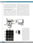
Figure 4. Tspan18 interacts with Orai1. (A) HEK-293T cells were transfected with a Myc- tagged human Orai1 expression construct and one of a panel of FLAG-tagged human tetraspanin constructs. Cells were lyzed in 1% digitonin and immunoprecipitated with an anti-FLAG antibody. Samples were separated by SDS-PAGE and both immunoprecipitated (IP) and whole cell lysate (WCL) samples were blotted with anti-FLAG and anti-Myc antibod- ies. A representative blot for each is shown (left) with quantitation of Myc-tagged Orai1 immunoprecipitated with the tetraspanins (right). Data were nomalized by logarithmic transformation before analysis by one-way ANOVA and Dunnett’s post test. Error bars rep- resent Standard Error of Mean (SEM) from three independent experiments. *P<0.05. (B) HeLa cells were transfected with Myc-tagged human Orai1, FLAG-tagged human Tspan18, or both constructs. Cells were fixed and stained with an anti-Myc antibody (green), an anti-FLAG antibody (red), and imaged by confo- cal microscopy (left). The Manders' coeffi- cients (M1 and M2) were calculated from the confocal stacks to quantify the degree of over- lap (right). Error bars represent the SEM from three independent experiments.
1898
haematologica | 2019; 104(9)
B
