
Page 222 - 2019_09-HaematologicaMondo-web
P. 222
P.J. Noy et al.
mine. Soluble vWF release, as detected by ELISA, was reduced by approximately 90% compared to control cells (Figure 7A). This was corroborated by immunofluores- cent staining of vWF that showed minimal release of Weibel-Palade bodies after Tspan18 knockdown (Figure 7B). Similar to Tspan18, Orai1 knockdown reduced Weibel-Palade body release after thrombin stimulation,
but knockdown of Orai2 or Orai3 had no effect (Figure 7C). Furthermore, Tspan18 knockdown reduced platelet adhesion to a thrombin-activated HUVEC monolayer by 85-90% (Figure 7D). These data support a role for Tspan18 and Orai1 in vWF release and platelet capture following endothelial cell activation by inflammatory mediators.
AC
D
FG
E
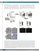
Figure 6. Tspan18-knockout mice have a hemostasis defect due to the absence of Tspan18 expression by non-hematopoietic cells. (A) Tail bleeding assays were performed by amputating 3 mm tail tips of anesthetized mice and the weight of blood lost was measured. The mice were Tspan18+/+, Tspan18-/-, or lethally irradiated Tspan18-/- or Tspan18+/+ mice reconstituted with fetal liver cells from Tspan18+/+ or Tspan18-/- embryos. Each symbol represents one animal. All data were analyzed by Fisher’s exact test. *P<0.05; ***P<0.001. Note that bleeding was stopped by cauterizing the tails of some mice, because of regulations limiting the amount of blood loss on our Home Office License. (B and C) Washed platelets from Tspan18+/+ or Tspan18-/- mice were activated with 3 μg/mL collagen (B) or 0.3 U/mL thrombin (C), and aggregation was measured by light transmission with stirring. Quantitated percentage aggregation each minute is shown. Error bars represent the Standard Error of Mean (SEM) from at least three pairs of litter-matched mice. (D and E) Plasma samples from Tspan18+/+ and Tspan18-/- mice were subjected to a prothrombin time test with human placental thromboplastin (D) and an activated partial thromboplastin time test with purified soy phosphatides with ellagic acid (E). Error bars represent SEM from four pairs of litter-matched mice. (F) Immunohistochemistry was used to show a grossly normal vasculature in Tspan18+/+ and Tspan18-/- mice formalin-fixed paraffin-embedded 5 μm sections from kidney and pancreas, using the MECA32 anti-mouse panendothelial cell antibody. Images are representative of three pairs of litter-matched mice. (G) Confocal microscopy was used to show a grossly normal vasculature in Tspan18+/+ and Tspan18-/- mice ears, by staining ante- rior ear tissue with biotinylated isolectin GS-IB4 glycoprotein followed by Alexa647-conjugated streptavidin. Images are representative of three pairs of litter-matched mice. ImageJ quantitation of 3 fields of view (500 x 500 pixels) per mouse showed a mean total vessel length of 11131±1271 pixels for Tspan18+/+ and 11684±283 pixels for Tspan18-/- (n=3; error represents SEM). mins: minutes.
1900
haematologica | 2019; 104(9)
B
