
Page 224 - 2019_09-HaematologicaMondo-web
P. 224
P.J. Noy et al.
A
C
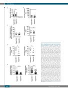
Figure 8. Tspan18-knockout mice have impaired histamine- D induced von Willebrand Factor (vWF) release and impaired thrombo-inflammatory responses. (A) Tspan18+/+ and Tspan18-/- mice were intraperitoneally-injected with hista- mine and plasma vWF levels were measured 30 minutes (min) later by ELISA. Error bars represent Standard Error of Mean (SEM) from eight Tspan18+/+ and seven Tspan18-/- mice. *P<0.05. (B) Tspan18+/+and Tspan18-/- mice were anesthetized, and the abdominal aorta exposed and mechanically injured through a single firm compression with forceps. Blood flow was subsequently monitored with a Doppler flowmeter to calculate the time until complete occlu- sion of the vessel. Each symbol represents one animal. (C) Tspan18+/+ and Tspan18-/- mice were anesthetized and the mesentery was exteriorized through an abdominal incision. Platelets were fluorescently labeled with Dylight 488-conju- gated anti-GPIX derivative. Small mesenteric arterioles were exposed to FeCl3-induced chemical injury via topical applica- tion. Time to appearance of the first thrombi was recorded (left), and the time until complete occlusion of the vessel was measured using fluorescence intravital microscopy (right). Each symbol represents one animal. (D) Tspan18+/+ and Tspan18-/- mice were anesthetized and surgery per- formed to stenose the inferior vena cava. After 48 hours (h), thrombus length (left) and weight (right) were measured. Each symbol represents one animal and horizontal bars rep- resent the median. *P<0.05. All data in (A-D) were analyzed by one-way ANOVA with Dunnett’s multiple comparisons test. (E) Myocardial ischemia-reperfusion injury was induced in the left ventricle of the beating heart of anesthetized mice by occluding the left anterior descending artery for 45 min with a suture. Reperfusion was instigated for 2 h by removal of the ligature, after which the organ was harvested. Frozen sections were analyzed for the presence of platelets in the microcirculation by immunofluorescence microscopy. Three litter-matched pairs of Tspan18+/+ and Tspan18-/- mice were used, with three sections per mouse and five images ana- lyzed per section. Each symbol represents one image. For each image, the integrated density value was calculated as a representation of the total number of platelets (left), and the average aggregate size was also calculated (right). Error bars represent the SEM and data were analyzed by Mann-
E
Whitney test. ****P<0.0001.
1902
haematologica | 2019; 104(9)
B
