
Page 218 - 2019_09-HaematologicaMondo-web
P. 218
P.J. Noy et al.
the lack of effective antibodies to Tspan18. To test for an interaction using co-immunoprecipitation, transfected HEK-293T cells were lyzed in 1% digitonin, a stringent detergent that has been used previously to identify tetraspanin-partner protein interactions.22,47 FLAG-tagged Tspan18 co-immunoprecipitated with Myc-tagged Orai1,
A
but five other control tetraspanins did not (Figure 4A). Moreover, Tspan18 and Orai1 co-localized when expressed in HeLa cells, at a level of approximately 90% pixel co-localization when assessed using the Manders’ coefficient (Figure 4B). These data suggest that Tspan18 interacts with Orai1.
CD
E
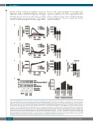
Figure 2. Tspan18-knockdown endothelial cells have impaired Ca2+ mobilization. (A-D) Human umbilical vein endothelial cells (HUVEC) were transfected with a neg- ative control siRNA (CON) or with one of two independent siRNA targeting Tspan18 (T18 KD). After 48 hours, HUVEC were loaded with the Ca2+-sensitive dye Fluo-4 NW and Ca2+ measurements taken using a FlexStation fluorescence reader during addition (arrow) of (A) 1 U/mL thrombin, (B) 20 μM histamine, or (C) 10 μM iono- mycin. Representative Ca2+ traces are shown for Tspan18-knockdown HUVEC (left), with quantitation of maximum intracellular Ca2+ concentrations (right). Data were analyzed by one-way ANOVA with Dunnett’s multiple comparisons test. Error bars represent the Standard Error of Mean (SEM) from three independent experiments. *P<0.05; ***P<0.001. (D) siRNA-transfected HUVEC from (A) to (C) were harvested, mRNA extracted, cDNA produced and Tspan18 mRNA levels were assessed by quantitative real-time polymerase chain reaction (qPCR). Data were normalized to 18S and actin as internal controls and adjusted such that the non-siRNA-trans- fected mock value was 1 in each experiment. Data were then normalized by logarithmic transformation, and analyzed by one-way ANOVA and Tukey’s multiple com- parison test. Error bars represent the Standard Error of the Mean from three independent experiments. ***P<0.001. (E) HUVEC were subjected to Tspan18 siRNA knockdown as described for (A-D), stimulated with 1 U/mL thrombin or 20 μM histamine for 5 minutes, then whole cell lysates were analyzed by western blotting with phospho-ERK1/2 and total ERK1/2 antibodies. (Left) Representative blots. (Right) Quantitation of three independent experiments. Error bars represent SEM. Knockdown efficiency was similar to that shown in (D) (data not shown). secs: seconds; RFU: relative fluorescence unit.
1896
haematologica | 2019; 104(9)
B
