Page 217 - 2019_09-HaematologicaMondo-web
P. 217
Tspan18 regulates Orai1 in endothelial cells
AB
CD
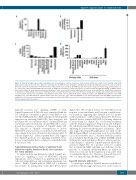
Figure 1. Tspan18 is highly expressed by endothelial cells. (A) Quantitative real-time polymerase chain reaction (qPCR) was carried out for Tspan18 using cDNA derived from a panel of mouse tissues. Data were normalized for the HPRT housekeeping gene and adjusted such that the lung value was 100 in each experiment. Error bars represent Standard Error of Mean from three independent tissue samples. (B) RNA-Seq data from major cell types in mouse lung, generated by Du et al.,40 was used to show Tspan18 mRNA expression levels as fragments per kilobase of transcript sequence per million mapped fragments (FPKM). (C) RNA-Seq data from major cell types in mouse brain, generated by Zhang et al.,41 was used to show Tspan18 mRNA expression levels as described in (B). (D) qPCR was carried out for Tspan18 on cDNA derived from a panel of primary human cells [dermal fibroblasts, aortic smooth muscle, hepatocytes, peripheral blood leukocytes from buffy coat and human umbilical vein endothelial cells (HUVEC)], and human cell lines (HEK-293T human embryonic kidney cells, MDA-MB-231 epithelial cells, DAMI megakaryocytic cells, HEL and K562 erythroleukemia cells, U937 monocytic cells, Jurkat and HPB-ALL T cells, DG75 and Raji B cells and HMEC-1 microvascular endothelial cells). Data were normalized for actin and adjusted such that the HUVEC value was 100 in each experiment. Error bars represent Standard Error of Mean from two independent cell samples.
Tspan18 activates Ca2+ signaling, MAPK or both, Tspan18-transfected DT40 cells were stimulated with the Ca2+ ionophore ionomycin or phorbol ester PMA to acti- vate the MAPK pathway. PMA synergized with Tspan18 expression in activating NFAT/AP-1, but ionomycin did not (Figure 3B). As a positive control, combined PMA and ionomycin induced relatively strong NFAT/AP-1 activa- tion in the presence or absence of Tspan18 (Figure 3B). The capacity of Tspan18 overexpression to induce NFAT/AP-1 activation was not restricted to DT40 B cells, since similar data were obtained in the human Jurkat T- cell line (Figure 3C). Taken together, these data suggest that Tspan18 promotes Ca2+ signaling and NFAT activa- tion via a mechanism that is common to endothelial cells, B cells and T cells.
Tspan18-induced nuclear factor of activated T-cell activation requires functional Orai1 store-operated Ca2+ entrychannels
To understand the mechanism by which Tspan18 pro- motes Ca2+ signaling, a series of NFAT/AP-1 reporter experiments were conducted in gene-knockout DT40 cells and using inhibitors and a dominant-interfering con- struct. Firstly, Tspan18-induced NFAT/AP-1 activation was found to be independent of the three IP3 receptors
(Figure 3D). IP3 receptors release Ca2+ from ER stores in response to tyrosine kinase and G protein-coupled recep- tor activation, suggesting that Tspan18 does not operate on these pathways or IP3 receptors themselves. However, Tspan18 did not activate NFAT/AP-1 following chelation of extracellular Ca2+ (Figure 3E), or following treatment with the immunosuppressive drug cyclosporin A (Figure 3F), which prevents NFAT translocation to the nucleus by inhibiting its activatory phosphatase calcineurin. These data suggest that Tspan18 might induce Ca2+ entry via the SOCE channel Orai1, a major entry route for extracellular Ca2+ in non-excitable cells.8 Consistent with this possibil- ity, a dominant interfering form of Orai1 (E106Q), which multimerizes with endogenous Orai1 to yield a non-func- tional channel,44-46 inhibited Tspan18-induced NFAT/AP-1 activation (Figure 3G). As a positive control to confirm that downstream NFAT signaling was still intact in the presence of dominant interfering Orai1, its inhibitory effect was overcome by the expression of an active form of calcineurin (Figure 3G). Therefore, Tspan18 may acti- vate Ca2+ entry through the Orai1 SOCE pathway.
Tspan18 interacts with Orai1
To investigate whether Tspan18 interacts with Orai1, transfected epitope-tagged proteins were used because of
haematologica | 2019; 104(9)
1895


