
Page 219 - 2019_09-HaematologicaMondo-web
P. 219
Tspan18 regulates Orai1 in endothelial cells
Orai1-knockdown endothelial cells have impaired Ca2+ mobilization and Orai1 surface expression requires Tspan18
To determine whether knockdown of Orai1 could phe- nocopy Tspan18 knockdown, intracellular Ca2+ mobiliza- tion was measured following siRNA-mediated knock- down of Orai1. This resulted in impaired Ca2+ mobiliza- tion in response to thrombin (Figure 5A) or histamine (Figure 5B). As a control, knockdown of the other Orai family members, Orai2 and Orai3, did not affect Ca2+ mobilization (Figure 5A and B), in agreement with previ- ous studies on Orai proteins in HUVEC.48,49 Positive con- trol ionomycin treatment gave a sustained intracellular Ca2+ response in all samples (Figure 5C) and effective
A
CD
knockdown was confirmed by qPCR (Figure 5D-F).
A common mechanism of tetraspanin function is to regulate their partner proteins by facilitating their exit from the endoplasmic reticulum (ER) and trafficking to the cell surface.1,50,51 To determine whether Orai1 localiza- tion could be regulated by Tspan18 in this manner, HUVEC were lentivirally transduced with Myc-Orai1 and transfected with control or Tspan18 siRNA duplexes, and Orai1 subcellular localization assessed by confocal microscopy. Orai1 was localized primarily to the cell periphery in control cells, but this was reduced following Tspan18 knockdown, with Orai1 partially co-localized with the ER marker calnexin (Figure 5G). Quantitative analyses showed that approximately 40% of Orai1 was
EFG
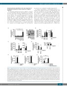
Figure 3. Tspan18 overexpression in cell lines activates Ca2+-responsive NFAT signaling in an Orai1-dependant manner. (A) The DT40 B cell line was transfected with an NFAT/AP-1-luciferase reporter construct, a β-galactosidase expression construct driven by the elongation factor (EF)-1a promoter to control for transfection efficiency, and FLAG-tagged mouse tetraspanin constructs or empty vector control. At 24 hours (h) post transfection, cells were lyzed and assayed for luciferase and β-galactosidase. Luciferase data were normalized for β-galactosidase values (left). Whole cell lysates from these cells were separated by SDS-PAGE and blotted with an anti-FLAG antibody. (Right) Representative blot. (B and C) The DT40 B cell line (B) and the human Jurkat T cell line (C) were transfected with an NFAT/AP-1- luciferase reporter construct and β-galactosidase expression construct with (+) or without (-) FLAG-tagged mouse Tspan18. At 24 h post transfection cells were stim- ulated for 6 h with 50 ng/mL PMA or 1 μM ionomycin (Iono) (left), or both together (right). Luciferase assays were then performed as described in (A). (D) DT40 cells with (+) or without (-) expression of FLAG-tagged mouse Tspan18 were tested for NFAT/AP-1 luciferase activity as described in (A), but using cells with gene knockouts of the three IP3 receptors (IP3R-) in comparison to wild-type (WT) cells (left). Whole cell lysates were western blotted with an anti-FLAG antibody (right). (E and F) DT40 cells with (+) or without (-) expression of FLAG-tagged mouse Tspan18 were tested for NFAT/AP-1 luciferase activity as described in (A), except that cells were treated with 4 mM EGTA as a Ca2+ chelator (E) or with 2 μM cyclosporin A as a calcineurin inhibitor (F). (G) DT40 cells were transfected with FLAG-tagged human Tspan18 in the presence or absence of a dominant negative human Orai1 E106Q mutant construct, or a consitutively active human calcineurin construct. The exper- iment was conducted as described for (A). All luciferase data were corrected for β-galactosidase values, normalized by logarithmic transformation, and analyzed by one-way ANOVA and Tukey’s multiple comparison test. *P<0.05; **P<0.01; ***P<0.001. Error bars represent the Standard Error of the Mean from at least three independent experiments. ns: not significant.
haematologica | 2019; 104(9)
1897
B
