
Page 110 - 2019_08-Haematologica-web
P. 110
S. Liu et al.
KLF4 and RUNX1 was abrogated by RUNX1-ETO in a dose-dependent manner (Figure 5C), suggesting that RUNX1-ETO competed with RUNX1 for binding to KLF4 and abrogated transcription of KLF4.
Biological roles of RUNX1 and KLF4 in t(8;21) leukemia cells
The results seen in Figure 1 suggested that RUNX1 and KLF4 might contribute to the apoptosis and differentiation of Kasumi-1 cells induced by PB. Overexpression experi- ments were then performed to address their roles in t(8;21) leukemia cells. RUNX1 and KLF4 were over- expressed respectively in Kasumi-1 cells by a pCDH
lentivirus system, and the overexpression efficiencies were confirmed at both mRNA and protein levels by qRT- PCR and western blot assay (Figure 6A). 3-(4,5- dimethylthiazol-2-yl)-5-(3-carboxymethoxyphenyl)-2-(4- sulfophenyl)-2H-tetrazolium (MTS) assay was performed to evaluate their effects on cell proliferation. The results showed that both of them could markedly inhibit cell pro- liferation (Figure 6B). Cell cycle analysis was carried out with propidium iodide staining and examined by flow cytometry. KLF4 overexpression blocked cell cycle in G0/G1 phase and reduced S and G2/M cell proportion, while overexpression of RUNX1 had no significant effect on the cell cycle in Kasumi-1 cells (Figure 6C). Apoptosis
A
B
C
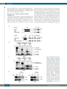
Figure 4. Identification of the specific domains of RUNX1 and RUNX1-ETO mediating interaction with KLF4. (A) Schematic representation of full-length and truncation mutants of RUNX1 (top) and RUNX1-ETO (bottom). (B and C) FLAG-KLF4 and full-length or truncation mutants of RUNX1 (B) or RUNX1-ETO (C) with MYC tag were co-transfected into 293T cells. At 48 hours (h) after transfec- tion, cell lysates were prepared and underwent immunoprecipitation (IP) with anti-FLAG antibody. Immunoblotting (IB) analyses were performed with anti-MYC antibody. Different combinations of co- transfection with KLF4 and full-length or truncation mutants of RUNX1/RUNX1- ETO were labeled as KLF4/RUNX1, KLF4/RUNX1-ΔRHD, KLF4/RHD, KLF4/RE, KLF4/RE-ΔRHD and KLF4/RHD, respectively. (D) KLF4 and ETO expression plasmids were co-trans- fected into 293T cells. At 48 h after transfection, cells were harvested and cell lysates underwent IP with anti-KLF4 or anti-ETO antibody. IB analysis was per- formed with the other antibody. WB: western blotting.
D
1602
haematologica | 2019; 104(8)
