
Page 111 - 2019_08-Haematologica-web
P. 111
RUNX1 transactivated and interacted with KLF4
analysis demonstrated that both RUNX1 and KLF4 pro- moted apoptosis of Kasumi-1 cells (Figure 6D). Further evidence of cell apoptosis was provided by Wright- Giemsa staining, which displayed typical apoptotic char- acteristics such as karyopyknosis, reduced ratio of nucleus to cytoplasm, nuclear fragmentation with intact cell mem- brane, and vacuolar degeneration in pCDH-RUNX1 and pCDH-KLF4 groups compared with that of the control group (Figure 6E). We next examined the effects of RUNX1 and KLF4 overexpression on Kasumi-1 cell differ- entiation. The expression levels of myeloid cell surface markers CD11b and CD15 were analyzed by flow cytom- etry at 48 h and 72 h after lentivirus infection. The results showed that there was a big increase in the proportion of CD11b+ and CD15+ cells after infection with pCDH-KLF4, whereas no parallel changes were found in pCDH-RUNX1 group (Figure 6F and Online Supplementary Figure S3).
Identification of P57 as a target gene of KLF4
As mentioned in Figure 1, the upregulation of P57 corre- lated with that of RUNX1 and KLF4 during PB-induced cell differentiation and apoptosis; however, the mecha- nism of this coherence was unknown. Previously pub- lished studies reported that KLF4 could up-regulate P57 in lymphatic endothelial cells and colon cancer cells.18,19 Based on this, we explored whether KLF4 up-regulated the expression of P57 in t(8;21) leukemia cells. Gene promoter analysis showed that there were four KLF4 putative bind- ing sites (K1~K4) within P57 promoter region (bp -979 to +240) (Homo sapiens chromosome 11, GRCh38) (Figure
7A). We then cloned the promoter region of P57 contain- ing KLF4 putative binding sites into a pGL3-Basic Vector to construct the pGL3-P57 plasmid. Next, the pGL3-P57 plasmid was co-transfected with different doses of KLF4 expression plasmid into CV-1 cells and the luciferase activ- ity of pGL3-P57 was evaluated. KLF4 could transactivate pGL3-P57 in a dose-dependent manner with the maximal effect with an almost 8-fold increase at the dose of 100 ng (Figure 7B). Moreover, P57 protein level was found remarkably up-regulated in Kasumi-1 cells after KLF4 overexpression, which further substantiated the regula- tion relationship (Figure 7C). As discussed (see above), KLF4 was a downstream gene of RUNX1; we therefore concluded that RUNX1, KLF4 and P57 might make up a transcriptional activation cascade in t(8;21) leukemia cells. To further testify the “RUNX1-KLF4-P57” pathway, we over-expressed RUNX1 in Kasumi-1 cells and examined the expression level of putative downstream genes KLF4 and P57. The result showed that both KLF4 and P57 were remarkably up-regulated after RUNX1 overexpression (Figure 7D). Finally, we explored the biological functions of P57 in t(8;21) leukemia cells. Overexpression of P57 mediated inhibition in cell proliferation, blockage in cell cycle, and induction of cell apoptosis, similar to RUNX1 and KLF4 (Figure 7E). However, P57 overexpression had no effects on cell differentiation as assessed by myeloid markers CD11b and CD15 and morphology analysis (Figure 7E and Online Supplementary Figure S3). Taken together, the above results suggested that the “RUNX1- KLF4-P57” pathway, activated by PB, was closely related to cell proliferation and apoptosis in t(8;21) leukemia cells.
ABC
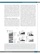
Figure 5. RUNX1-ETO competes with RUNX1 for binding to KLF4. (A) Fixed dose of pCMV5-FLAG-KLF4 and pCMV5-MYC-RUNX1 were co-transfected with increasing dosages of pCMV5-MYC-RUNX1-ETO into 293T cells. At 48 hours (h) after transfection, cell lysates were prepared and underwent immunoprecipitation (IP) with anti- FLAG antibody. Immunoblotting analyses were performed with anti-MYC antibody. β-actin and H3 served as loading controls for the whole cell lysate. (B) The effects of RUNX1 and RUNX1-ETO on KLF4-dependent transactivation of target genes. CV-1 cells were transfected with fixed dose of KLF4 target genes reporter plasmid (KLF4-Reporter), pCMV5-KLF4 and increasing doses of pCMV5-RUNX1 (top) or pCMV5-RUNX1-ETO (bottom). At 48 h after transfection, transfected cells were har- vested for luciferase assay. The luciferase transcriptional activities of KLF4-Reporter were measured and normalized to that of Renilla luciferase. Cells transfected with only KLF4-Reporter were set as control. (C) The transcriptional regulatory effects of RUNX1 and RUNX1-ETO on KLF4 target gene P57 promoter reporter (pGL3- P57). CV-1 cells were transfected with KLF4 or RUNX1 expression plasmid alone or different combinations of KLF4, RUNX1 and RUNX1-ETO expression plasmids together to investigate their regulating effect on pGL3-P57. At 48 h after transfection, transfected cells were harvested for luciferase assay and western blot analysis. The luciferase transcriptional activities of pGL3-P57 were measured and normalized to that of Renilla luciferase. Cells transfected with pGL3-P57 only were set as control.
haematologica | 2019; 104(8)
1603
