
Page 109 - 2019_08-Haematologica-web
P. 109
RUNX1 transactivated and interacted with KLF4
ETO-mediated interaction with KLF4 were also investi- gated. The results showed that KLF4 not only interacted with RHD, but also with the ETO part of RUNX1-ETO (Figure 4A and C). Further study verified the physical interaction between KLF4 and ETO protein (Figure 4D).
RUNX1-ETO competes with RUNX1 for binding to KLF4
We investigated the physiological interaction between RUNX1 and KLF4. We found that both RUNX1 and RUNX1-ETO interacted with KLF4 (Figure 3) and leukemic fusion protein RUNX1-ETO obstructed the physiological interaction between RUNX1 and KLF4. Fixed doses of pCMV5-FLAG-KLF4 and pCMV5-MYC- RUNX1 were co-transfected with increasing doses of pCMV5-MYC-RUNX1-ETO into 293T cells. At 48 h after transfection, cell lysates were prepared for Co-IP assay. RUNX1-ETO competed with RUNX1 for binding to KLF4 in a dose-dependent manner (Figure 5A). To further inves- tigate the effects of RUNX1 and RUNX1-ETO on KLF4-
dependent transactivation of target genes, a luciferase reporter plasmid containing KLF4 binding sites (KLF4- Reporter) was constructed. Luciferase assay was then per- formed (Figure 5B). The results demonstrated that KLF4 was capable of transactivating its target genes reporter plasmid (KLF4-Reporter) in CV-1 cells. RUNX1 significant- ly enhanced the transcriptional activation effect of KLF4 in a dose-dependent manner, while RUNX1-ETO could only enhance the effect slightly at low doses (50-100 ng). The above results suggested that RUNX1-ETO might compete with RUNX1 for binding to KLF4 and abrogate transcrip- tion of the KLF4 target gene. To confirm this hypothesis, we performed co-transfection of RUNX1 and RUNX1- ETO for luciferase assay using KLF4 target gene P57 pro- moter reporter. CV-1 cells were transfected with KLF4 or RUNX1 expression plasmid alone or in different combina- tions of KLF4, RUNX1 and RUNX1-ETO expression plas- mids to investigate their regulating effect on P57 promoter reporter. The transactivation of P57 promoter mediated by
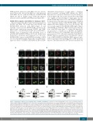
AB
CD
Figure 3. Identification of KLF4 as a novel binding partner of RUNX1 and RUNX1-ETO. (A) KLF4 co-localized with RUNX1 in nuclei. (Top) Co-localization analysis of exogenous MYC-RUNX1 and FLAG-KLF4 in 293T cells at 48 h after transfection by immunofluorescence assay. Anti-MYC (green) and anti-FLAG (red) antibodies were used as primary antibodies; DAPI (blue) was used for nuclear staining. Scale bar represents 10 μm. (Middle and bottom) Co-localization analysis of endogenous RUNX1 and KLF4 in Kasumi-1 and SKNO-1 cells with anti-RUNX1 (green) and anti-KLF4 (red) antibodies; DAPI (blue) was used for nuclear staining. Scale bar repre- sents 10 μm. (B) KLF4 co-localized with RUNX1-ETO in nuclei. (Top) Co-localization analysis of exogenous MYC-RUNX1-ETO and FLAG-KLF4 in 293T cells at 48 h after transfection. Antibodies were used as described in (A). Scale bar represents 10 μm. (Middle and bottom) Co-localization analysis of endogenous RUNX1-ETO and KLF4 in Kasumi-1 and SKNO-1 cells with anti-ETO (green) and anti-KLF4 (red) antibodies. DAPI (blue) was used for nuclear staining. Scale bar represents 10 μm. (C) KLF4 interacted with RUNX1. 293T cells were co-transfected with pCMV5-MYC-RUNX1 and pCMV5-FLAG-KLF4. At 48 h after transfection cells were harvested and cell lysates underwent immunoprecipitation (IP) with anti-FLAG or anti-MYC antibody. Immunoblotting (IB) analysis was performed with the other antibody. (D) KLF4 interacted with RUNX1-ETO. 293T cells were co-transfected with pCMV5-MYC-RUNX1-ETO and pCMV5-FLAG-KLF4. At 48 h after transfection, cells were harvested and cell lysates underwent IP with anti-FLAG or anti-MYC antibody. Immunoblotting analysis was performed with the other antibody. WB: western blotting.
haematologica | 2019; 104(8)
1601
