
Page 107 - 2019_08-Haematologica-web
P. 107
RUNX1 transactivated and interacted with KLF4
Results
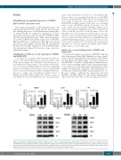
Phenylbutyrate up-regulated expressions of RUNX1, KLF4 and P57 in Kasumi-1 cells
Gene expression profiles of PB-treated Kasumi-1 cells demonstrated that RUNX1, KLF4 and P57 were up-regu- lated during the process of cell differentiation and apopto- sis induced by PB. To confirm the upregulation of these three genes, Kasumi-1 cells were treated with PB at differ- ent doses and incubation times. Then cells were harvest- ed, and the mRNA and protein expressions of RUNX1, KLF4 and P57 were evaluated by qRT-PCR and western blot. The results clearly showed that PB up-regulated RUNX1, KLF4 and P57 expressions at both mRNA (Figure 1A) and protein (Figure 1B) level in a time- and dose- dependent manner.
Identification of KLF4 as a novel target gene of RUNX1 and RUNX1-ETO
To determine the mechanism of KLF4 upregulation by PB treat- ment, we first analyzed KLF4 promoter region (bp -2668 to -195) (Homo sapiens chromosome 9, GRCh37) for the transcription fac- tors binding sites by Jaspar database. The result showed that there were seven putative RUNX1-binding sites (R1~R7) within KLF4 promoter region (Figure 2A). We then constructed a pGL3-KLF4 promoter reporter plasmid (pGL3-KLF4) containing RUNX1 bind- ing sites to analyze the transcriptional regulation effects of RUNX1 and RUNX1-ETO on KLF4. The luciferase activity of pGL3-KLF4 was activated by RUNX1 in a dose-dependent manner
with a nearly 12-fold increase at the dose of 200 ng (Figure 2B). However, there was a less than 3-fold increase of pGL3-KLF4 luciferase activity by RUNX1-ETO at the doses from 50 ng to 200 ng. To further determine the specific binding sites of RUNX1 and RUNX1-ETO on KLF4 promoter, ChIP assays were performed with a specific anti-RUNX1 antibody in Kasumi-1 cells. The results confirmed the direct binding of RUNX1 to KLF4 promoter region via four predicted binding sites at bp -2617 to -2607 (R1), bp -2312 to -2302 (R2), bp -1136 to -1126 (R4), and bp -764 to -754 (R5) (Figure 2C). Subsequently, western blot analyses were per- formed to verify the regulation relationship on protein level. 293T cells were transfected with pCMV5-vector, pCMV5-RUNX1 and pCMV5-RUNX1-ETO plasmids, respectively. After 48 h of trans- fection, cells were harvested for western blot. The results showed that KLF4 was clearly up-regulated by RUNX1 while RUNX1- ETO had almost no effect (Figure 2D).
KLF4 is also a novel binding partner of RUNX1 and RUNX1-ETO
De novo motif analysis of our previous RUNX1 Chip-Seq data demonstrated that KLF4 binding sites were signifi- cantly enriched within RUNX1 chip regions, which raised the hypothesis that KLF4 might co-localize and interact with RUNX1. To validate our hypothesis, immunofluores- cence confocal imaging analysis and co-immunoprecipita- tion (Co-IP) assays were performed. Immunofluorescence analysis showed both exogenous and endogenous co- localization of RUNX1 and KLF4 in nuclei (Figure 3A). Co- IP assay further confirmed the interaction between RUNX1 and KLF4 (Figure 3C and Online Supplementary Figure S1A). However, due to the low expression level of
A
B
Figure 1. Upregulation of RUNX1, KLF4 and P57 in t(8;21) leukemia cells by sodium phenylbutyrate (PB) treatment. (A) Relative expressions of RUNX1, KLF4 and P57 mRNA were measured by quantitative real-time polymerase chain reaction in Kasumi-1 cells after treatment with different doses of PB for the time (hours, h) indicated. The gene transcript levels were normalized to those of GAPDH and set to 1 in the control group. (B) Western blot analysis of RUNX1, KLF4 and P57 protein expressions in Kasumi-1 cells after PB treatment. β-actin and H3 were used as internal loading controls.
haematologica | 2019; 104(8)
1599
