
Page 108 - 2019_08-Haematologica-web
P. 108
S. Liu et al.
KLF4 in Kasumi-1 and SKNO-1 cells, the endogenous interaction between KLF4 and RUNX1 was less evident than that under conditions of exogenous overexpression in 293T cells. The co-localization and protein interaction of fusion protein RUNX1-ETO and KLF4 were also inves- tigated. Similar to wild-type RUNX1, RUNX1-ETO co- localized and interacted with KLF4 in nuclei (Figure 3B and D and Online Supplementary Figure S1B).
Identification of the specific domains of RUNX1 and RUNX1-ETO mediating interaction with KLF4
A large number of studies have reported that the runt homology domain (RHD) was responsible for RUNX1
interacting with other transcription factors, such as PU.1, STAT5 and GATA1.3,4,17 To clarify whether RUNX1 inter- acted with KLF4 through RHD, full-length or truncated mutant (RHD deleted, ΔRHD) of RUNX1 and RHD domain were cloned into a pCMV5 MYC-tagged plasmid, respectively (Figure 4A). After co-transfecting 293T cells with each of the MYC-tagged RUNX1 constructs and FLAG-tagged KLF4 construct, Co-IP assays were per- formed with anti-FLAG antibody for IP and anti-MYC antibody for immunoblotting (IB). Both the full-length RUNX1 and RHD could directly interact with KLF4, while the truncated mutant RUNX1-ΔRHD failed to interact with KLF4 (Figure 4B). The specific domains of RUNX1-
A
B
C
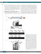
Figure 2. KLF4 is a novel target gene of RUNX1 and RUNX1-ETO. (A) Schematic representation of KLF4 pro- moter fragments fused to a pGL3-Basic Vector. The putative RUNX1 binding sites are indicated by orange boxes as follows: R1 (-2617 to -2607), R2 (-2312 to - 2302), R3 (-2014 to -2004), R4 (-1136 to -1126), R5 (- 764 to -754), R6 (-399 to -394), and R7 (-263 to -258). Transcription start site (TSS) is indicated by an arrow. Numbers represent base pairs relative to TSS. (B) pGL3- KLF4 promoter reporter plasmid (pGL3-KLF4) was co-
D transfected with increasing doses of pCMV5-RUNX1 or pCMV5-RUNX1-ETO into CV-1 cells. At 48 h after trans- fection, the luciferase transcriptional activity of pGL3- KLF4 was measured and normalized to that of Renilla luciferase. (C) Chromatin immunoprecipitation analysis of RUNX1 binding sites to KLF4 promoter region in Kasumi-1 cells. The predicted binding sites R1, R2, R4 and R5 were successfully amplified, both from the input DNA and chromatin immunoprecipitation by an anti- RUNX1 antibody, whereas no amplified product was obtained in the IgG control group. White stars indicate non-specific amplified bandings. (D) 293T cells were transfected with pCMV5-vector, pCMV5-RUNX1 and pCMV5-RUNX1-ETO, respectively. At 48 h after transfec- tion, cells were harvested and the protein levels of KLF4 were assayed by western blot. β-actin and H3 were used
as protein loading controls.
1600
haematologica | 2019; 104(8)
