
Page 199 - 2019_07 resto del Mondo-web
P. 199
Abnormal proplatelet formation in PT-VWD
(Figure 2B and Online Supplementary Figure S5A). PT-vWD megakaryocytes extended very long and branched pro- platelets on fibrinogen and vWF, probably due to the boosting role of bound vWF on proplatelet formation.9-11 However, the number of proplatelet tips was reduced and tip diameter was larger in PT-vWD megakaryocytes. In particular, we identified a subset of cells producing a num- ber of tips below the 95% Confidence Interval (95%CI) of
controls, and which had tips of a diameter above the 95%CI of controls (Figure 2C and D). Moreover, a signifi- cantly higher number of PT-vWD megakaryocytes extended proplatelets when plated on type I collagen, compared to control megakaryocytes (Figure 2B and E). Comparable results were obtained using mouse TgG233V megakaryocytes (Online Supplementary Figure S6).
In order to exclude the possibility that defects of the col-
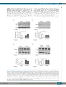
AB
CD
Figure 3. Collagen-triggered signaling in human megakaryocytes. (A) RhoA activation (RhoA-GTP) of megakaryocytes in suspension (-) or after 16 hours (h) of adhe- sion to type I collagen (+). Densitometric analysis was performed using the Image J software. Quantification of RhoA-GTP is relative to total RhoA expression and is expressed in arbitrary units (AU) (n=5, #Significantly different from control on type I collagen; P<0.05). (B) MLC-2 phosphorylation (pMLC-2) of megakaryocytes in suspension (-) or after 16 h of adhesion to type I collagen (+). Densitometric analysis was performed using Image J software. Quantification of pMLC-2 is relative to total MLC-2 expression and is expressed in arbitrary units (AU). (n=5; #Significantly decreased vs. control on type I collagen; P<0.05). (C) SFK phosphorylation (p-Src Tyr416) in megakaryocytes in suspension (-) or after 16 h of adhesion to type I collagen (+). Densitometric analysis was performed using Image J software. Quantification of p-SFK is relative to total SFK expression and is expressed in arbitrary units (AU) (n=5, *Significantly increased vs. control in suspension; P<0.05). (D) Lyn phosphorylation (p-Lyn) in megakaryocytes in suspension (-) or after 16 h of adhesion to type I collagen (+). Lyn was immunoprecipitated and western blotting was carried out using the anti-SFK antibody. Densitometric analysis was performed using Image J software. Quantification of P-Lyn is relative to total Lyn expression and is expressed in arbitrary units (AU) (n=5; *Significantly different from resting control; P<0.05). Blots are representative of megakaryocytes from five controls and five different preparations from the platelet-type von Willebrand disease patient.
haematologica | 2019; 104(7)
1477
