
Page 201 - 2019_07 resto del Mondo-web
P. 201
Abnormal proplatelet formation in PT-VWD
(Online Supplementary Figure S7). Interestingly, control megakaryocytes cultured in suspension showed phospho- rylation of Lyn when incubated with ristocetin, thus con- firming that the binding of vWF to GPIba triggers Lyn phosphorylation (Online Supplementary Figure S8A). Finally, Cofilin, a protein that is phosphorylated by activated RhoA,30 was less phosphorylated upon adhesion to colla- gen in PT-vWD than in control megakaryocytes (Online Supplementary Figure S8B).
Platelets in bone marrow
Immunohistochemistry showed an increased number of platelets in the bone marrow of our PT-vWD patient com- pared to healthy controls (Figure 4A). In order to exclude the possibility that the increased number of platelets was simply due to the enhanced number of megakaryocytes in bone marrow (Online Supplementary Figure S1), we count- ed platelets in bone marrow from patients with ITP who also show increased megakaryocytes in bone marrow, but here the number of platelets was comparable to controls (Online Supplementary Figure S9A). Similarly, bone marrow from TgG233V mice showed an increased number of platelets compared to bone marrow from TgWT mice (Figure 4B). The ratio between bone marrow platelets and circulating platelets, as assessed by flow cytometry, was also signifi- cantly increased in TgG233V mice (Online Supplementary Figure S9B).
Platelet life span and platelet-bound vWF
The number of circulating platelets was 572.3±32.6x10⁹/L in TgWT mice and 180.1±47.2x10⁹/L in TgG233V mice. TgG233Vmice showed a reduced platelet half-
life (26 hours) compared to TgWT mice (47 h). A significant- ly lower percentage of DyLight 488-stained platelets was observed already 24 h after fluorescent anti-GPIX mAb injection, and the difference became even more evident 48 h after, when residual DyLight 488-stained platelets in blood were 12% in TgG233V and 46% in TgWT mice (Figure 4C). Moreover, 71.3±12.2% and 83.2±14.6% of circulat- ing human and murine PT-vWD platelets showed surface- bound vWF, respectively, while no vWF could be detected on the platelet surface in controls. vWF bridged-platelet aggregates were also seen in PT-vWD, and not observed in controls (Figure 4D).
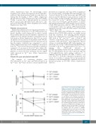
These data suggest that vWF/platelet complexes circu- lating in blood in PT-vWD patients are rapidly cleared from the circulation, thus reducing platelet lifespan. Indeed, the administration of DDAVP increased plasma vWF both in TgWT and TgG233V mice (+97±21% and +93±12%, respectively), but it caused a drastic reduction of platelet count only in TgG233V mice (Figure 5A). The per- centage of circulating DyLight 488-stained platelets was reduced by 30% in TgG233Vmice 60 min after DDAVP injec- tion while it did not vary significantly in TgWT mice, con- firming that vWF-bound platelets in PT-vWD are rapidly cleared from the circulation (Figure 5B). PT-vWD platelets did not show increased exposure of phosphatidylserine under resting conditions (% of Annexin V-positive platelets: PT-VWD, 4.1±0.6%, con- trols, 4.6±2.7%). From the comparative quantitative eval- uation of our studies, it emerges that ectopic proplatelet formation is the mechanism that contributes the most to thrombocytopenia in PT-vWD (Online Supplementary Table S2).
A
Figure 5. Effect of desmopressin (DDAVP) admin- istration on platelet count and lifespan in mice. (A) Platelet count was assessed by flow cytometry in saline-injected (circles) or DDAVP-injected (trian- gles) mice. A significant drop in platelet count was evident in TgG233V mice 30 minutes after the injec-
B tion of DDAVP. Circulating platelets are expressed as percentage of the platelets measured at time=0. Data represent mean±Standard Error of Mean (SEM) (n=5, *P<0.05 vs. TgWT). (B) Mice were infused with a DyLight 488-conjugated anti- GPIX mAb. Residual green fluorescent platelets were quantified by flow cytometry in saline-inject- ed (circles) or DDAVP-injected (triangles) mice. A significant drop in the percentage of fluorescent platelets was evident in TgG233V mice, but not in TgWT, 60 minutes after the injection of DDAVP. Circulating platelets are expressed as percentage of GPIX-positive platelets relative to the total CD41/61-PE positive platelet population meas- ured at time=0. Data represent mean±SEM (n=5;
*P<0.05 vs. TgWT).
haematologica | 2019; 104(7)
1479
