
Page 188 - 2019_07 resto del Mondo-web
P. 188
R. van Oorschot et al.
derived megakaryocytic cells were all members of the minichromosome maintenance complex (DNA replication licensing factor proteins, MCM2-7) (Online Supplementary Figure S6A, D) and structural maintenance of chromosome proteins (SMC1A, SMC2-4, SMCHD) (Online Supplementary Table S1), which are required for DNA repli- cation and chromosome condensation, respectively (Figure 5D; Online Supplementary Table S2). Furthermore, MEIS1, a
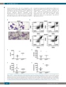
AB
key player in megakaryocyte differentiation which is nor- mally downregulated in the final stages of megakaryocyte differentiation,30 showed consistent expression in GFI1BQ287* iPSC-derived megakaryocytic cells while it was never detected in control iPSC-derived cells (Online Supplementary Figure S6C). Thus, the GFI1BQ287* protein influences the expression of a large number of proteins dur- ing megakaryopoiesis.
CD
EF
Figure 4. GFI1BQ287* of induced pluripotent stem cell-derived megakaryoid cells phenocopy disease characteristics. (A) Cytospins of control and GFI1BQ287* induced pluripotent stem cell (iPSC) lines differentiated towards megakaryocytes (MK) and stained with May-Grünwald Giemsa. Pictures were taken at 40x magnification using a Zeiss Scope.A1 microscope (Zeiss) and images were processed with Zen blue edition. (B) Representative flow-cytometric analysis of control and GFI1BQ287* iPSC-derived cells for surface expression of megakaryocyte-associated markers CD34, CD41a and CD42b. (C-E) Normalized median fluorescence intensity (nMFI) of CD34 (C), CD42b (D) and CD41a (E). (F) Quantification of CD41a+ megakaryocytic cells per seeded iPSC to measure expansion potential. CD41a+ cells were negative for erythroid, myeloid and endothelial makers indicating that these cells represent true megakaryocytic cells (data not shown) **P<0.01, ****P<0.0001.
1466
haematologica | 2019; 104(7)
