
Page 187 - 2019_07 resto del Mondo-web
P. 187
Molecular characterization of GFI1BQ287* mutation
but not detectable in GFI1BQ287* iPSC-derived cells (Online Supplementary Figure S6B). Other proteins related to inter- feron signaling, i.e. STAT1, IFI16, IFI30, IFI35, IFIT1, IFIT3, OAS2 and OAS3, were also significantly reduced in GFI1BQ287* iPSC-derived megakaryocytic cells compared to controls (Online Supplementary Table S1). Because the con- trol and GFI1BQ287* lines are derived from different individ- uals, differences in individual protein expression might be
A
B
caused by variation in genetic makeup of the lines. We, therefore, analyzed gene ontology (GO) terms to identify multiple differentially expressed proteins assigned to spe- cific pathways/functions. This identified downregulated proteins related to diverse functions, including mitochon- dria, response to stimuli, and transmembrane transport (Figure 5C; Online Supplementary Table S2). On the other hand, strongly enriched proteins in the GFI1BQ287* iPSC-
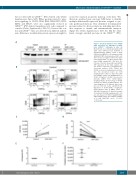
Figure 3. Chemical disruption of the GFI1B- LSD1 interaction by GSK-LSD1 recapitu- lates GFI1BQ287* hallmarks in vitro. (A) Western blot on co-immunoprecipitated (IP) LSD1 after GFP-trap bead pulldown in dimethylsulfoxide (DMSO) control or GSK- LSD1-treated MEG-01 cells transduced with GFI1B-GFP (WT). Non-transduced (NT) cells were used as a negative control. The upper panel shows LSD1 (~90 kDa) and the lower panel GFI1B variants-GFP (~58 kDa). The left side of the blot shows LSD1 and GFI1B- GFP expression in the input samples. (B) Cell surface expression of megakaryocyte- associated markers CD34, CD41a, and CD42b, to measure megakaryocyte matura- tion. The presented results are from megakaryocytic cultures 2 days after addi- tion of DMSO control or 4 μM GSK-LSD1. (C) CD34 normalized median fluorescence intensity (nMFI) from CD34+/CD41a+ megakaryoblasts. (D) Percentage of CD42b positivity on CD41a+ megakaryoblasts. The connecting lines (C-D) indicate which sam- ples are from the same experiment. (E) Expansion of CD34+/CD41a+ megakaryo- blasts during 2 days of DMSO control or GSK-LSD1 treatment. (F) Absolute number of proplatelet-forming megakaryocytic cells per view (based on Online Supplementary Figure S4A,B) 6 days after addition of DMSO or 4 μM GSK-LSD1 (n=6). *P<0.05, ***P<0.001.
CD
EF
haematologica | 2019; 104(7)
1465
