
Page 155 - 2019_07 resto del Mondo-web
P. 155
BCL2 limits crizotinib efficiency in ALk+ ALCL
MiR-34a-mediated BCL2 downregulation potentiates the antitumoral effects of crizotinib in NOD/SCID mice xenografted with ALK-positive anaplastic large cell lymphoma cells
Finally, we explored whether the combination of BCL2 downregulation and crizotinib treatment would have a significant therapeutic benefit in vivo. To address this ques- tion, NOD/SCID mice were xenografted with KARPAS- 299 cells that were either transfected with miR-Neg or miR-34a mimics. Crizotinib was administered orally for 22 days, during which time tumor growth was monitored (Figure 6A). On the day of sacrifice, tumors were weighed (Figure 6B) and samples were retrieved for immunohisto- chemistry (IHC) analysis (Figure 6C).
In accordance with the data collected in vitro, we observed that mice xenografted with miR-34a-transfected cells developed significantly smaller tumors than those xenografted with miR-Neg-transfected cells in the presence of crizotinib (Figure 6A and B). As seen in our in vitro viability assays, miR-34a-mediated BCL2 knockdown alone impaired tumor growth, albeit to a lesser extent than with the miR-34a/crizotinib combination. Hematoxylin & Eosin (HE) staining performed on samples excised from tumors treated with the miR-34a/crizotinib
combination also exhibited hallmarks of higher cell fragility (Figure 6C). To confirm our in vitro findings showing higher levels and deleterious effects of autophagy in KARPAS-299 cells under miR-34a/crizotinib combination, we looked at in vivo autophagy activity by performing LC3B and p62 IHC analyses in tissues from the tumor xenografts (Figure 6C and Online Supplementary Figure S12), as previously reported.37,38 We observed that dot-like patterns of both LC3B and p62 staining increased strongly and significantly in cells that received the combined miR-34a/crizotinib treatment in comparison with the single treatment. These results are consistent with increased autophagy activity.
Discussion
Our study is the first to report that the expression of ALK and BCL2, two major oncogenes, are inversely corre- lated in ALK-positive ALCL through an ALK-dependent BCL2 repression mechanism. Indeed, we demonstrated that BCL2 levels increased following either the pharmaco- logical inhibition (crizotinib treatment) or the siRNA-tar- geted knockdown of ALK (siALK transfection) in ALK-
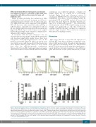
A
BC
Figure 3. BCL2 downregulation enhances crizotinib-triggered autophagic flux. (A) Flow cytometry analysis of autophagic flux following the knockdown of BCL2 and crizotinib treatment in KARPAS-299 cells expressing a tandem fluorescently-tagged LC3 reporter protein. mRFP-EGFP-LC3 KARPAS-299 cells (described in Online Supplementary Methods) were transfected with either negative controls (siCTL or miR-Neg) or with siBCL2 or miR-34a mimics. Twenty-four hours (h) later, transfected cells were treated or not with increasing doses of crizotinib (0 to 500 nM) for a further 48 h. Induction of autophagic flux was analyzed by monitoring the RFP/EGFP fluorescence ratio in individual cells. Cells were split into two groups based on their relative RFP/ EGFP fluorescence ratios: cells with low/basal autophagic flux and cells with high/induced autophagic flux. A representative experiment is shown. (B and C) Histograms representing the percentage of cells with a RFP/EGFP ratio reflective of high autophagic flux, from n=5 (siBCL2) or n=3 (miR-34a mimics) independent experiments ± Standard Error of Mean; *P≤0.05; **P≤0.01; ***P≤0.001; unpaired Student’s t-test.
haematologica | 2019; 104(7)
1433
