
Page 156 - 2019_07 resto del Mondo-web
P. 156
A. Torossian et al.
positive ALCL cells. These data are consistent with and complete previous studies reporting a lack of BCL2 pro- tein expression in primary tissue samples of ALK-positive ALCL31,32 and low mRNA levels in the ALK-positive ALCL cell line, KARPAS-299.39 This observation in a cancerous environment seems paradoxical but is balanced by the fact
that these tumor cells over-express MCL1, through a molecular mechanism that involves miR-29a.32,39,40 In the present study, to rule out a potential compensation of BCL2 downregulation by BCL2 isoforms in crizotinib- treated cells, we checked the expression of MCL1, BCL-XL/S and BCL-W and did not find any significant
A
B
CD
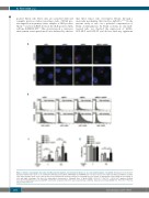
Figure 4. Enhanced autophagic flux induced by BCL2 downregulation and crizotinib treatment is associated with impaired cell viability. Twenty-four hours (h) after ULK1 knockdown either alone or in combination with BCL2 knockdown, mRFP-EGFP-LC3 KARPAS-299 cells were treated or not with crizotinib (200nM) for a further 72 h. Representative data of (A) confocal microscopy and (B) flow cytometry analysis of autophagic flux are shown. (C) Histograms representing the percentage of cells with high autophagic flux from five independent experiments ± Standard Error of Mean (SEM); *P≤0.05; **P≤0.01; ***P≤0.001; unpaired Student t-test. (D) Cell viability was assessed by MTS colorimetric measurements in the same experimental conditions. Data represent mean±SEM; n=3; *P≤0.05; **P≤0.01; unpaired Student t-test.
1434
haematologica | 2019; 104(7)
