
Page 154 - 2019_07 resto del Mondo-web
P. 154
A. Torossian et al.
mRNA (siULK1) and/or against BCL2 mRNA (siBCL2), in the presence or absence of crizotinib. ULK1 knockdown (validated by western blot analyses, Online Supplementary Figure S11) efficiently blocked the autophagy process, as revealed by a decrease in both yellow (reflecting autophagosomes) and red (reflecting autolysosomes) punctate staining (Figure 4A). Flow cytometry monitoring of autophagic flux confirmed firstly that the blockade of BCL2 expression increased the number of cells exhibiting high autophagy in both untreated and crizotinib-treated cells (Figure 4B and C), and secondly that ULK1 downregulation successfully blocked the autophagic machinery as it reduced basal autophagy levels and restrained the crizotinib-triggered autophagic flux observed in BCL2-knocked down cells (2-fold decrease). Most interestingly, MTS viability assays showed that the effects of BCL2 downregulation in further reducing cell viability in crizotinib-treated cells was completely reversed by the knockdown of ULK1 (Figure 4D). These results strongly suggest that BCL2 downregulation follow- ing crizotinib treatment reinforced autophagic flux, which was deleterious for ALK-positive ALCL cells, and thereby led to a greater loss in viability under these conditions.
To address the question of whether potentiation of the autophagic flux in our model was sufficient to trigger cell
death, we next performed experiments with a combination of crizotinib and rapamycin, a well-known mTOR inhibitor and strong inducer of autophagy, namely in ALK-positive tumor cells.35,36 We first confirmed that rapamycin alone (100 nM) did induce autophagy in KARPAS-299 cells as 60% and 75% of cells harbored a high autophagic flux following 24 h and 48 h of treatment, respectively (Figure 5A). We then observed that combining crizotinib and rapamycin treatments resulted in a clear potentiation of the autophagic flux, with more pronounced effects obtained with the lowest dose of crizotinib used in our assays (125 nM). We also found that this potentiation of the autophagic flux was associated with a decrease in cell viability, as measured by MTS col- orimetric assays (Figure 5B), an effect that was more pro- nounced after 48 h and 72 h of treatment. However, Annexin-V/PI staining revealed no significant differences in the number of cells undergoing apoptotic cell death when comparing crizotinib treatment alone with crizo- tinib and rapamycin combined treatment, independently of the dose of crizotinib used and the duration of the treat- ment (Figure 5C). Altogether these results indicate that excessive autophagy upon crizotinib and rapamycin co- treatment contributes to cell death independently of apop- tosis.
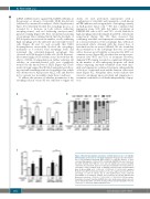
AB
C
Figure 2. BCL2 downregulation potentiates crizotinib-induced loss in cell viability, which involves an increase in cell death. Twenty-four hours (h) after the transfec- tion of BCL2-targeted interfering RNAs (siBCL-2 or miR-34a mimics), or their corresponding negative controls (siCTL or miR-Neg), KARPAS-299 were treated (or not) with crizotinib (500 nM) for 72 h. (A) Cell viability was assessed by MTS colorimetric measurement. Each set of data was normalized to its related untreat- ed negative control condition (siCTL or miR-Neg) and represents mean±Standard Error of Mean (SEM); n=3. **P≤0.01; ***P≤0.001; ****P≤0.0001; unpaired Student t-test. (B) Flow cytometry analysis of cell cycle. Graph represents the mean percentage of cells in sub-G1, G1, S and G2/M phases. Data represent mean±SEM; n=3; Statistical analysis was performed by two-way ANOVA with Bonferroni correction; **P≤0.01; ***P≤0.001; ****P≤0.0001. (C) Flow cytome- try analysis of annexin V-positive KARPAS-299 cells. Graph represents the percent- age of annexin V-positive cells from six independent experiments±SEM. *P≤0.05; **P≤0.01; ***P≤0.001; unpaired Student t-test.
1432
haematologica | 2019; 104(7)
