
Page 67 - 2019_04-Haematologica-web
P. 67
Impaired CVR in adults with sickle cell disease
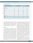
Table 3. Spearman’s correlation coefficients between clinical parameters and GM cerebral blood flow and GM cerebrovascular reserve in adult patients with sickle cell disease.
Parameter
Age
Baseline GM CBF Hemoglobin concentration ASAT
ALAT
Leukocytes
Platelets
HbF%
HbS%
MCV
Creatinine
CRP
Ferritin
Markers of hemolysis
Bilirubin
LDH Reticulocyte %
GM Cerebral blood flow (CBF) GM Cerebrovascular reserve (CVR) Spearman’s rho (ρ) P Spearman’s rho (ρ) P
-0.36 0.03 0.17 0.33
- -
- 0.61 <0.001* 0.26 0.17
- 0.03 0.85 0.30 0.09 0.30 0.42
- 0.41 <0.05 0.10 0.64 0.05 0.80 0.07 0.69 -0.08 0.71 0.11 0.57
0.43 0.01 0.45 0.01 0.32 0.06
- 0.43 0.01 0.40 0.02 0.01 0.94 0.12 0.49
- 0.18 0.32 - 0.41 0.03 0.37 0.07 - 0.32 0.13 -0.15 0.38
0.34
0.02 0.92
-0.18 0.35
-0.23 0.19 -0.18 0.33 -0.32 0.06
<0.05
*P values that remained significant after Benjamini-Hochberg procedure for multiple-comparison adjustment. GM CBF: granulocyte-macrophage cerebral blood flow; CVR: cere- brovascular reserve; ASAT: aspartate aminotransferase; ALAT: alanine aminotransferase; MCV: mean corpuscular volume; CRP: c-reactive protein; LDH: lactate dehydrogenase;
contour overlay in the bottom row of Figure 4. Binary logistic regression with lesion presence or absence as an outcome variable, showed that GM CVR was not a pre- dictor of lesion prevalence (P=0.28), and neither was WM CVR (P=0.57). The same was true for GM CBF (P=0.18), age (P=0.24), hemoglobin levels (P=0.08), and the other blood markers. In linear regression analysis, CVR was not a significant predictor of lesion volume (P=0.669).
Discussion
Chronic inflammation and hemolysis play a key role in the pathologic processes that can diminish the dilatory capacity of small resistance arteries in SCD. In our study, we observed a globally reduced gray matter CVR in patients with SCD without a history of stroke, compared to race-matched healthy controls. We found that CVR was particularly impaired in patients with high baseline CBF, indicating that in these patients, cerebral vasodilation was almost maximal at rest. Indeed, the lowest CVR of 5% was observed in a patient with SCD and comorbid moyamoya syndrome, in whom the highest CBF and dif- fuse white matter injury was observed. Silent cerebral infarcts (SCIs) were detected in the majority of patients, but these were not related to any of the hemodynamic MRI markers. The association between CBF and elevated lactate dehydrogenase and bilirubin levels suggest that blood flow may be related to higher hemolytic rate. However, this association was not significant after adjust- ment for multiple comparisons so remains to be investi- gated in future studies.
Acetazolamide was well-tolerated by all participants
and did not induce vaso-occlusive crisis in any of the patients, indicating that this test can be performed safely to assess CVR in patients with SCD. A previous study also using intravenous acetazolamide administration in chil- dren with SCD to assess CVR with SPECT16 did not report on safety of acetazolamide, so it remains unclear if the authors had the same experiences regarding side-effects. Acetazolamide has the advantage over CO2 inhalation of inducing maximal dilation without metabolic changes, which allows a true assessment of vasodilatory capacity.
We observed a plateau in the response to acetazolamide after 10-15 minutes, corresponding to maximal vasodila- tion. However, there was a difference in the maximal CBF between the groups, clearly showing a reduced vascular reserve capacity. This difference in CVR can be explained by chronically increased resting vessel diameter, as we have shown previously,21 which leaves these patients with little reserve for further vasodilation. Numerous resting ASL studies in children with SCD have shown that chron- ic anemia leads to high CBF20,29–32 and the high flow requirements in SCD could lead to a loss of autoregulatory capacity if dilatory reserve is being used for perfusion. Our dynamic CBF response supports our hypothesis that adult patients with SCD have severely reduced vasodilatory capacity. Hence, autoregulatory capacity is being used to maintain basal cerebral oxygenation, posing a risk for cerebral ischemia and infarction.
The prevalence of SCIs found in our cohort was 81%, which is in line with previous reports on lesions in adults with SCD ranging from 15% to 90%.9,26–28,33 The large aforementioned variation arises from methodologic differ- ences including improvements in imaging technology pro- viding higher sensitivity, differences in age between
haematologica | 2019; 104(4)
695
