
Page 69 - 2019_04-Haematologica-web
P. 69
Impaired CVR in adults with sickle cell disease
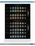
Figure 4. Axial slices of cohort averaged registered maps of hemodynamic MRI parameters. Upper panel shows CBF at baseline and post acetazolamide in GM for healthy controls and patients with sickle cell disease (SCD), and indicates clearly that CBF is elevated in SCD patients in all brain regions. Middle panel shows cohort- averaged co-registered CVR maps in GM and WM and illustrates that CVR is lower in patients with SCD compared to healthy controls in both grey and white matter regions of interest. CVR was not uniformly distributed and appeared to be higher in posterior compared to anterior regions in healthy controls and appear to be higher in watershed and periventricular regions in patients with SCD, with lowest CVR appearing in the deep white matter interface with grey matter. Bottom panel shows the lesion contours overlaid on white matter CVR in patients with SCD. CBF: cerebral blood flow; CVR: cerebrovascular reserve; GM: gray matter.
haematologica | 2019; 104(4)
697
