
Page 68 - 2019_04-Haematologica-web
P. 68
L. Václavu° et al.
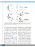
AC
B
D
E
Figure 3. Differences between patients and controls, and associations among gray matter CBF, gray matter CVR, and hemoglobin. (A) The boxplot shows higher CBF in gray matter (GM) in patients with sickle cell disease (SCD) compared to healthy controls. (B) CVR in GM in patients with SCD was half of that of controls. (C) The scatterplot shows the significant association between CBF in GM and hemoglobin concentration in SCD patients. (D) The scatterplot shows that CVR in GM was significantly associated with hemoglobin levels in SCD. (E) The magnitude of the CVR in GM was significantly associated with resting CBF in GM. CBF: cerebral blood flow; CVR: cerebrovascular reserve; GM: gray matter; SCD: sickle cell disease.
patient cohorts,28 the size and number of the lesions,34 as well as differentiation between silent cerebral infarcts and lacunar infarcts.3,26 This sensitivity to technology was pre- viously demonstrated in a study by our group performed in adult SCD patients,28 in which a 7T imaging field strength with 0.8 mm isotropic resolution was compared to 3T with 1 mm isotropic resolution, and found a lesion prevalence of 50% at 3T and 90% at 7T field strength, respectively. The relatively high prevalence of lesions (45%) identified in the control group seems consistent with having higher detection sensitivity when using improved technology.28 Hence, a consensus on lesion def- inition and measurement that is harmonized across tech- nologies and sites is needed in order to compare studies from several cohorts in future studies.
As demonstrated in the lesion density map in our study, SCI prone areas were primarily located in the deep white matter, watershed and borderzone regions,35 where perfu- sion is known to be lower, and ischemic risk thought to be higher in children with SCD,36 than in the cerebral cortex. While the pathogenesis of SCIs in SCD is still unclear, some evidence does show that the severity of anemia plays a role in SCIs in children with SCD.37 Even though insufficient cerebral oxygen delivery and the associated ischemic risk is probably due to chronic anemia, recent work demonstrated that additional acute moments of crit- ical hypoperfusion may lead to lesions rather than chronic anemia by itself.7,22 Limited cerebrovascular reserve may
be the underlying condition that places patients at increased vulnerability for inadequate oxygen delivery and extraction during acute anemia or superimposed hypoxia. Hence, low CVR itself may not be a sufficient condition to initiate lesion formation, but an additional crucial ‘second hit’, such as acute anemic events or super- imposed hypoxia, is probably necessary. We hypothesize that interventions that improve oxygen delivery such as blood transfusion, hydroxyurea or new disease-modifying drugs that reduce hemolysis may improve CVR and there- by reduce risk for cerebral ischemia and infarction. Whether reduced CVR is an independent risk factor for SCIs remains to be demonstrated in a prospective trial.
The limitations of this study include potential selection bias of patients with no history of stroke. This may have induced a bias towards less severe patients and thereby also less severe white matter injury burden. Indeed, lesions were mostly small punctate lesions, with a total median lesion volume of 0.34 mL, which is low compared to total brain volume. However, since these lesions were present in the majority of patients, our cohort is likely to be a reasonable representation of the adult SCD popula- tion without overt stroke. We included 36 patients with SCD under the premise that we would have enough power to detect differences in CVR between patients and healthy controls based on an a priori sample size calcula- tion. However, given that the correlation between lesion volume and CVR has not been studied previously in SCD,
696
haematologica | 2019; 104(4)
