
Page 54 - 2019_04-Haematologica-web
P. 54
C.J. Mercadante et al.
which may reflect chronic effects of iron excess and/or anemia. Overall, given that body iron levels would not be affected by iron redistribution between organs, we hypothesized that the long-term change in body iron lev- els in transferrin-treated Trfhpx/hpx mice reflected changes in absorption and/or excretion.
Trf+/+ and treated Trfhpx/hpx mice have similar 59Fe absorption rates
Untreated Trfhpx/hpx mice absorb iron excessively.14 If transferrin treatment suppresses absorption rates below excretion rates, body iron levels would decrease without any need for increased excretion rates in Trfhpx/hpx mice. To test this hypothesis, we performed intragastric 59Fe gav- age in 2.5-month old Trf+/+, untreated Trfhpx/hpx, and transfer- rin-treated Trfhpx/hpx mice, then analyzed 59Fe levels 16 h later. From herein we studied males and females separate- ly to detect sex-specific differences. Untreated Trfhpx/hpx mice absorbed more gavaged 59Fe than did Trf+/+ and treat- ed Trfhpx/hpx mice, and Trf+/+ and treated Trfhpx/hpx mice absorbed the same amount of 59Fe (Figure 4A). Similar results were observed when absorption values were nor- malized to body mass (Figure 4B) or when mice were analyzed 1 h after gavage (data not shown). This indicated that reversal of iron excess in transferrin-treated Trfhpx/hpx mice was not due to ‘hypersuppression’ of iron absorp- tion.
Trf+/+ and treated Trfhpx/hpx mice have similar 59Fe half-lives
We next assessed excretion in Trf+/+, untreated Trfhpx/hpx, and transferrin-treated Trfhpx/hpx mice using 59Fe. Mice from the absorption studies were used, as we rationalized that gavage was the most physiological means of administer- ing 59Fe. We repeatedly measured body 59Fe levels in mice for 2 months from 2.5 to 4.5 months of age (Figure 4C).
We ended the excretion study at 4.5 months as untreated Trfhpx/hpx mice do not consistently survive past this age. Body 59Fe levels, plotted versus time, were fitted to expo- nential decay curves. Exponential decay equations were then used to calculate two factors: biological 59Fe half- lives, expressed in days, and percent body 59Fe excreted per day, referred to here as ‘59Fe excretion rates’. 59Fe half- lives and 59Fe excretion rates are inversely proportional to each other. 59Fe half-lives were ~80-120 days in all mice except for untreated male Trfhpx/hpx mice, which had a half- life of ~170 days (Figure 4D). 59Fe excretion rates were ~0.6-0.8% in all mice except for male Trfhpx/hpx mice, which had an excretion rate of ~0.45% (Figure 4E). Notably, 59Fe half-lives and excretion rates did not differ between Trf+/+ and untreated Trfhpx/hpx mice.
Trf mice excrete iron largely via the gastrointestinal tract
During the 2-month excretion study, mice were placed repeatedly in metabolic cages for overnight collections of feces and urine. Fecal and urinary 59Fe levels were expressed as a percent of body 59Fe levels at the time of collection, then averaged for each mouse group. Most 59Fe was excreted in feces (Figure 4F). Body 59Fe losses could be accounted for by fecal and urinary 59Fe losses in all mice except transferrin-treated Trfhpx/hpx females (Figure 4G). Overall, these data indicate that Trf mice excreted iron largely via the gastrointestinal tract.
59Fe-based analyses predict relative abundance of urinary iron and fecal ferritin in Trf mice
In some of the earliest radioisotope-based studies of iron excretion in mice, Finch and colleagues multiplied body 59Fe excretion rates by body iron levels to estimate the amount of iron excreted per day.17,18 We employed this approach here to further explore iron excretion in Trf
ABC
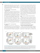
Figure 3. Long-term transferrin treatment corrects body iron excess in Trfhpx/hpx mice. (A-C) Body masses (A) and iron (Fe) levels (B) and concentrations (C), measured in Trf+/+ mice (‘+/+’, orange) and untreated Trfhpx/hpx mice (‘hpx/hpx’, green) from 1 to 6 months of age and in Trfhpx/hpx mice treated with transferrin (TF) from 2 to 6 months of age (‘hpx/hpx +TF’, blue). Top and bottom graphs differ only by markers of significance (P<0.05) assessed by two-way analysis of variance with a Holm- Sidak post-hoc test. In top panels, for a given age, different letters indicate values that differ significantly. In bottom panels, for a given group, the letter indicates that a value differs significantly from the 2-month old value. In all panels, dashed lines indicate that only two untreated Trfhpx/hpx mice survived to 6 months. Data are represented as mean ± standard error of mean: each value represents data from five mice, with males and females grouped together.
682
haematologica | 2019; 104(4)
