
Page 53 - 2019_04-Haematologica-web
P. 53
Iron excretion in iron excess
(see Online Supplement for details). Based upon these calcu- lations, it is likely that changes in tissue iron levels in the first 2 weeks of transferrin treatment largely represent mobilization of iron to the bone marrow.
To explore the effect of transferrin treatment on body iron levels in Trfhpx/hpx mice beyond the first 2 weeks of treatment, we measured body iron content in 1- to 6- month old Trf+/+ and Trfhpx/hpx mice and in Trfhpx/hpxmice treat- ed with transferrin from 2 to 6 months of age. The mice were euthanized in order to measure body iron content and no blood was removed prior to euthanasia. After mouse pelts had been removed, gastrointestinal tracts were isolated and cleared of contents. Pelts, cleared gas-
AB
C
trointestinal tracts, and carcasses were then analyzed for iron levels, which were summed to calculate body iron levels. We focused on body iron levels here as these would not be affected by redistribution between organs. Untreated Trfhpx/hpx mice were smaller than Trf+/+ mice, and treatment increased body sizes of Trfhpx/hpx mice (Figure 3A). Body iron levels (in mg iron) and concentrations (in μg iron/g body mass) were greater in untreated Trfhpx/hpx mice than in Trf+/+ mice (Figure 3B,C). Treatment of Trfhpx/hpx mice resulted in no difference in iron levels and a less than two-fold difference in iron concentrations relative to those in Trf+/+ mice by 6 months of age (Figure 3B,C). Most untreated Trfhpx/hpx mice did not survive to 6 months,
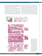
Figure 2. Trfhpx/hpx mice accu- mulate iron in the duodenum. (A-C) Mice from Figure 1 were analyzed for duodenal iron (Fe) levels by inductively cou- pled plasma absorption emis- sion spectrometry (A) and tis- sue Fe staining in duodenal smooth muscle (B) and villi (C). In (A), data are represent- ed as the mean ± standard error of mean. Brackets indi- cate statistical significance (P<0.05) calculated by one- way analysis of variance with a Holm-Sidak post-hoc test. Each value represents data from five mice, with males and females grouped together. In (C), the arrowhead indicates detectable Fe staining in duo- denal enterocytes.
haematologica | 2019; 104(4)
681
