
Page 52 - 2019_04-Haematologica-web
P. 52
C.J. Mercadante et al.
Long-term transferrin treatment corrects body iron excess in Trfhpx/hpx mice
Several scenarios could explain decreased iron concen- trations in organs of transferrin-treated Trfhpx/hpx mice. Given the severity of anemia in untreated Trfhpx/hpxmice, the increase in hemoglobin levels in treated mutant mice
should require mobilization of a significant amount of iron from the liver and other organs to the bone marrow. We estimated that 0.30 mg of iron were mobilized from the liver and pancreas during the first 2 weeks of treat- ment and that the anemia in untreated mutant mice cor- responded to a deficit of 0.475 mg of hemoglobin iron
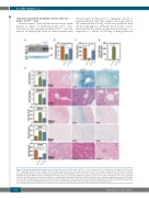
ABCD
E
Figure 1. Short-term transferrin treatment reduces tissue iron excess in Trfhpx/hpx mice. (A-D) Trf+/+ (‘+/+’), untreated Trfhpx/hpx (‘hpx/hpx’), and transferrin (TF)-treated Trfhpx/hpx (‘hpx/hpx +TF’) mice were analyzed at 2.5 months of age. Treated mice were injected with TF from 2 to 2.5 months of age. (A) Serum TF levels, measured by immunoblot (top) and Coomassie-stained protein gel (bottom). (B) Hemoglobin levels, measured by complete blood count. (C) Plasma hepcidin levels, measured by ELISA. (D) Splenic RNA level ratios of Fam132b to Actb (β-actin), measured by quantitative polymerase chain reaction and normalized to Trf+/+ levels. (E) Organ iron (Fe) levels (left panels), measured by inductively coupled plasma absorption emission spectrometry, and tissue Fe distribution (right micrographs), assessed by tissue Fe stain. In (B-E), data are represented as the mean ± standard error of mean. Brackets indicate statistical significance (P<0.05) calculated by one-way analy- sis of variance with a Holm-Sidak post-hoc test. Each value represents data from five mice, with males and females grouped together.
680
haematologica | 2019; 104(4)
