
Page 79 - 2019_02-Haematologica-web
P. 79
NGS for genetic diagnosis of AML
QiaSeqTM panels, respectively (Online Supplementary Figure S8B). Therefore, amplicon sequencing enables variant detection with analytical sensitivities that, in well-covered regions, are equivalent to those of Sanger sequencing, the reference method for clinical mutation testing.34,35
Larger DNA sequence variants such as FLT3-ITD and KMT2A-PTD require special attention in NGS data analy- sis as they may be missed by common variant calling tools.36,37 Thus, previous authors have added ITD-seek to the TruSight® Myeloid analysis pipeline.7 Applying this tool to our MV4-11 dilution series, we identified a 34 bp insertion in FLT3 exon 14, which closely matched pub- lished results,38 with an analytical sensitivity of 10% (Online Supplementary Figure S9A). The FLT3-ITD was not detected in the 10% MV4-11 sample using the QiaSeqTM panel with smCounter analysis, presumably because of suboptimal coverage achieved in our sequencing runs. However, given that our work with commercial kits aimed at clarifying their principal applicability for diagnostic pur- poses, we did not repeat these experiments.
For the identification of KMT2A-PTD in amplicon sequencing data, we developed PTDi by adapting a tool that had been used previously with a capture-based target- ed sequencing approach.36 PTDi analysis revealed amplifi- cation of exons 3-8 in patient AML-7 with a known e3e9 KMT2A-PTD (Online Supplementary Figure S9B, Online
Supplementary Table S9). Thus, not only are short DNA variants detectable by amplicon sequencing, but also diffi- cult ITD and PTD. Taken together, our results clearly con- firm that combining RNA- and DNA-based amplicon pan- els allows all major translocations and all types of clinically relevant mutations in AML to be uncovered by NGS.
Evaluation of the comprehensive next-generation sequencing platform for the diagnosis of acute myeloid leukemia in a clinical setting
After testing the performance of all sequencing modules and bioinformatics procedures, we evaluated the clinical utility of our platform as a diagnostic tool. As our kary- otype studies in cell lines clearly showed that NGS detects a higher number of numerical aberrations than chromo- some banding (Online Supplementary Tables S5 and S6), we first investigated a potential need for manual review of the raw CAI[N] output in order to avoid overestimation of karyotype complexity and enable appropriate risk stratifi- cation (Table 1, Online Supplementary Tables S10 and S11). We performed lc-WGS on additional patients' samples and reconstructed CNV karyotypes by cross comparison of CAI[N] results and known cytogenetic findings. All non- complex karyotypes, including three samples with pre- sumably normal karyotypes in which cytogenetic analysis had failed, were identified correctly (AML-5, -7-11a/b, -13,
A
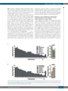
B
Figure 3. Detection of whole chromosome gains and losses by copy number variation karyotyping. Whole genome libraries from (A) an individual with Down syn- drome (T21) and (B) the BEN-MEN-1 cell line were sequenced with low coverage and analyzed by CAI[N]. RF: random female (N=2,819), RM: random male (N=2,605). Error bars represent the standard deviation (below visibility).
haematologica | 2019; 104(2)
283
