
Page 81 - 2019_02-Haematologica-web
P. 81
NGS for genetic diagnosis of AML
A
B
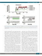
Figure 5. Sensitivity of copy number variation karyotyping. Genomic DNA from the BEN-MEN-1 cell line (monosomy 22) was diluted in healthy donor DNA (Healthy F1, Figure 2) in different ratios and subjected to lc-WGS and CAI[N] analysis. (A) Region plots for chromosome 22. The range ±3 standard deviations around the mean is indicated in pale red. (B) CNV decision plots. Read numbers in 1 Mb windows were normalized to 1x10⁶ total reads. RF: random female (N=2,819). Color coding in (B) as in (A).
benchtop sequencing device and commercially available reagents and kits, thus facilitating implementation even at smaller diagnostic centers that currently cannot offer the full spectrum of molecular and cytogenetic analyses required for a complete genetic work-up of AML. NGS results differ from diagnostic reports obtained by current standard techniques in two major aspects: first, NGS kary- otyping by lc-WGS and fusion transcript analysis does not reveal insights into the potential clonal heterogeneity of the disease, and second, DNA-based mutation testing yields variant allele frequencies rather than allelic ratios. While the observed frequency of cytogenetic abnormalities in a certain number of metaphases is not relevant for risk stratification, mutational burden is highly prognostically relevant for FLT3-ITD. Allelic ratios calculated from frag- ment analysis are not equivalent to variant allele frequen- cies, but based on our data presented here we cannot pro- pose a conversion from variant allele frequencies to the established risk stratification parameter.
In the method described here, sequencing resources are most effectively reduced by fusion detection on the level of RNA using anchored multiplex PCR for target enrichment. Anchored multiplex PCR requires previous knowledge on only one fusion partner, which is targeted by a gene-specif- ic primer included in the sequencing panel,10 so that even novel translocations involving commonly rearranged genes can be detected using a relatively low-complexity primer pool. Moreover, if a translocation does not result in a fusion transcript, but rather in transcriptional activation of
a target gene, such as MECOM overexpression in AML with inv(3)(q21.3;q26.2) or t(3;3)(q21.3;q26.2),44 the under- lying chromosomal rearrangement may be deduced from relative expression analyses via molecular barcode quan- tification. In contrast, translocation detection on the level of DNA needs to cover large intronic breakpoint regions and therefore requires higher throughput sequencing equipment or limitation of the assay to a subset of AML- relevant translocations.40,45,46
Most importantly, our approach provides an easy to use tool for “numerical” karyotyping, that – differently from single nucleotide polymorphism-karyotyping methods40 – enables CNV analysis in a completely unbiased manner without the need for specific capture probes. Notably, the CAI[N] algorithm does not uncover absolute ploidy, but classifies gains and losses even in strongly altered hypo- or hyperdiploid cases (e.g. HL-60 and NB-4 cells).
Despite mapping to chromosome bands, CNV kary- otyping by lc-WGS and CAI[N] analysis identifies chro- mosomal gains and losses at higher resolution than con- ventional cytogenetics (1 Mb versus 5-10 Mb). Subcytogenetic CNV have been observed previously by array-based comparative genomic hybridization in both complex and normal karyotype AML, including recurrent aberrations with potential prognostic impact, such as gain of 8q24.32,41,47 Moreover, NGS karyotyping does not require short-term culture of the sample material and therefore eliminates a major technical challenge of classi- cal cytogenetics and potential biases in clonally heteroge-
haematologica | 2019; 104(2)
285
