
Page 80 - 2019_02-Haematologica-web
P. 80
E.K.M. Mack et al.
A
B
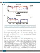
Figure 4. Detection of partial chromosome losses and gains by copy number variation karyotyping. Whole genome libraries from three AML patients’ samples were sequenced with low coverage and analyzed by CAI[N]. (A) Region plots for chromosome 5. (B) Region plot of chromosome 1 for patient AML-1. Read numbers in 1 Mb windows were normalized to 1 x10⁶ total reads. RF: random female (n=2,819), RM: random male (n=2,605). See also Online Supplementary Figures S1-S3.
-16, -17, -19, HES-1 / AML-14, -15, -20). In two samples that had not been characterized extensively by FISH, CNV karyotyping apparently identified marker chromosomes or detected additional aberrations (AML-2 / AML-1). Moreover, CAI[N] revealed copy number changes for five of six chromosomes involved in translocations in a highly complex karyotype (AML-3), but missed loss of chromo- some 10, which had been detected in <10% of cells by interphase FISH. In four patients, for whom cytogenetics had not been performed when samples were taken, NGS recovered at least a subset of aberrant clones or a normal karyotype as reported at initial diagnosis (AML-4, -6, -12 / AML-18, respectively). Taken together, overall karyotype complexity was determined correctly in all cases and 13/13 samples without risk-defining translocations were accu- rately assigned to prognostic groups based on CNV kary- otyping alone (Online Supplementary Table S10). Thus, we did not specifically validate discordant results between conventional and NGS karyotyping.
Next, to study patients’ samples in an unbiased manner, a group of four of the authors performed blinded analysis of CNV, fusion genes and mutations on eight additional AML samples and one acute lymphocytic leukemia sample (AML-21–28, ALL-1;39 Table 1, Online Supplementary Tables S7-S9). Two samples were excluded before unblinding because of insufficient read coverage resulting from poor DNA quality (AML-26, -27). In the remaining samples, CNV karyotyping uncovered all expected numerical
changes and one additional aberration, gain of chromo- some 19 in patient AML-28. Fusion analysis identified all translocations previously described in these patients. Variant analysis revealed no single nucleotide variants, insertions, deletions or ITD in this series of samples, in agreement with reference results. These findings further underscore the potential diagnostic value of our assay for the clinical management of AML.
Discussion
Here we developed and validated an integrated diagnos- tic platform that exclusively utilizes state-of-the-art NGS technology to obtain comprehensive clinically relevant insights into AML genomes. Our targeted approach covers all genetic features that define subclasses of AML with recurrent genetic abnormalities and/or prognostic groups as well as potentially actionable mutations2,9 and thus can serve as a potential alternative for diverse classical cytoge- netics and molecular biology assays in certain laboratory settings. In contrast to a previously described single-run NGS assay for AML diagnosis,40 we did not include detec- tion of copy number neutral loss of heterozygosity,41,42 as its prognostic value in AML43 is not fully established.2
We examine numerical aberrations, translocations, short sequence variants, FLT3-ITD and KMT2A-PTD in a rapid, robust and reliable composite assay. Our platform uses a
284
haematologica | 2019; 104(2)
