
Page 78 - 2019_02-Haematologica-web
P. 78
E.K.M. Mack et al.
22, which is found in all BEN-MEN-1 cells. These findings indicate that the sensitivity of CAI[N]-CNV karyotyping is sufficient to detect highly prevalent chromosomal aberra- tions in AML samples with a blast count of at least 20% without prior enrichment of the blast population.
Fusion gene and DNA variant detection
Our composite assay relies on predesigned amplicon panels for the detection of fusion transcripts and DNA vari- ants. Amplification strategies implemented in these kits10 or, respectively, specific panels, have already been exten- sively evaluated.7,28 Thus, we focused our studies on the detection of all subclass-defining translocations and all major types of clinically relevant DNA variants in adult AML. To investigate coverage of important fusion genes, we analyzed cell lines and patients’ samples harboring or lacking frequent chimeric transcripts. All expected fusions were identified in KASUMI-1,29 ME-1,30 NB-4,21 AML-5, AML-6 and CML-1, including two variants31 of BCR-ABL1 in the last sample. On the other hand, no fusions were detected in HL-6019 cells and in a patient with hypere-
A
osinophilic syndrome (HES-1), as reported by the reference laboratory (Online Supplementary Figure S8A, Online Supplementary Table S7). In a pool of the four cell lines, all fusions were recovered, but in a 1:25 dilution thereof, only the RUNX1-RUNX1T1 fusion transcript was identified. This finding underlines that RNA-based fusion detection is expression-dependent, so that the sensitivity of the assay varies for different samples and fusions.
Moreover, we exemplarily tested the TruSight® Myeloid panel (Illumina) and the QIASeqTM Myeloid Neoplasms panel (Qiagen), which incorporates molecular barcodes for PCR-error correction,32 as screening tools to identify short DNA variants in AML genomes. All single nucleotide vari- ants detected in HL-60, NB-4, ME-1, MV4-11 and SKNO-1 cells by the TruSight® Myeloid panel were consistent with COSMIC33 data or confirmed by Sanger sequencing, and the QiaSeqTM panel uncovered all reported mutations in samples from two patients with AML (Online Supplementary Table S8). Sequencing a dilution series of MV4-11 DNA revealed detection limits for the two p53 mutations of 1% and 10% with the TruSight® and
B
C
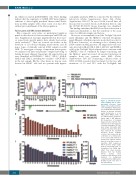
Figure 2. Calculated chromo- some banding and in silico- generated reference kary- otypes. (A) Calculated chromo- some banding by CAI[N] analy- sis of lc-WGS data. Read distri- butions in genomic windows along chromosome 9 of an AML patient (AML-2, Table 1) are indicated (left: cytogenetic bands, right: 1 Mb windows. (B) Frequencies of uniquely mapped reads on whole chro- mosomes for in silico-generat- ed normal karyotypes. RF: ran- dom female (N=2,819), RM: random male (N=2,605). Error bars represent the standard deviation (below visibility; <0.01%). Note that in (A) the centromere of a chromosome is not covered and in (B) the Y chromosome appears smaller than its actual size because of repetitive DNA sequences, which prevent unique align- ment of sequencing reads. (C) Scalability of the CAI[N] algo- rithm: Four whole genome libraries from two healthy female donors were sequenced with different read numbers in multiplexed sequencing runs (right panel). Healthy F1.1-4: four runs of the same library.
282
haematologica | 2019; 104(2)
