
Page 77 - 2019_02-Haematologica-web
P. 77
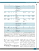
NGS for genetic diagnosis of AML
continued from the previous page
AML-185 45 M5
AML-19 68 M3v
AML-20 61 n.a
AML-21 18 M2
AML-22 31 M5
AML-23 48 M4eo
AML-24 41 RAEB-t
AML-25 57 M1
AML-26 72 M0/M1
AML-27 66 M4
AML-28 54 M5a
CML-1 74 CML
HES-1 73 HES
PB
BM
BM
PB
BM
PB
BM
PB BM
BM
BM
PB
BM
89% 97%
80% 90%
100% 98%
n.a
46XY, t(15;17)(q22;q12)[2]; 46XY,t(1;6;7) (p36;p21;q22), t(15;17)(q22;q12)[11]
n.a
(FLT3-ITD, NPM1_D, DNMT3A R882H, IDH1 R132H)
46,XX,
del(7q),t(8;21)(q22;q22)
48-49,XY,
+19, +der(21)X1-2[cp4]; 46,XY[7]
46,XY [6];
46,XY, inv. (16)(p13q22) [18]
45,XY, -7 [7],
46, XY, -7 +21 [9], 46, XY [5]
46,XX [20]
nuc ish 5p15(hTERTx3), 5q31(CDC25C,EGR1x3), 8cen(D8Z2x4), 17p13(TP53x3), 17q11(NF1x3)
45,XY,-7[9];
45,XY,-7.ish del(12)(p13p13) (3'ETV6-)[3];
45,XY,-7.ish del(12)(p13p13) (3'ETV6-,5'ETV6-)[6], 46,XY[4]
46,XY,t(9;11)(p22;q23)
46,XX,
t(9;22)(q34;q11)[20]
46, XY [15]
47,XX,
t(1;9)(q22;q34),+17 [3], 46,XX,t(1;9)(q22;q34),der(6) t(6;17)(p23;q21)[2]
46,XY
46,XY
46,XY
46,XX,del(7)(q33q36.2)
48,XY,+19,+21
46,XY
46,XY,-7,+21
46,XX
poor DNA quality; evidence for del(5p),
del(5)(q31), add(8p), +17
poor DNA quality/ too few reads; evidence for, -7,
del(12p13.2)
47,XY,+19
n.d
46,XY
n.d.
2,037,251
2,241,454
3,603,346
1,104,847
2,042,269
1,940,532
1,661,351
2,326,960 920,209
590,092
867,272
n.a.
2,072,387
n.a.
1,604,338
1,772,271
,2838,440
875,403
1,602,693
1,523,373
1,297,049
1,848,467 643,682
472,009
687,922
n.a.
1,636,629
n.a.
ALL-1 62 Common BM B-ALL
n.a.
n.a.
80%
10%
70% n.a.
n.a.
n.a.
n.a.
n.a.
80%
>30%
58%
63%
11%
60% n.a.
n.a.
n.a.
n.a.
n.a.
70%
1SampleAML-1:chromosome15wasnotcoveredbyFISHanalysesperformedbythereferencelaboratory(probesincludedWCP1/WCP18,WCP7,EGR1,WCP5,ATM,p53,IGHBCL2). 2Sample AML-4: cytogenetic analyses were not performed at the time the sample was taken for NGS karyotyping: 12 months earlier (BM): 45,X,der(X;7)(q10;p10)[7]; 46,XX,add(7)(q21)[6], 46,XX [3], 46,XY[4]. nuc ish Xp11.1-q11.1(DXZ1x2),Yq12(DYZ1x0)[50]/ Xp11.1-q11.1(DXZ1x1),Yq12(DYZx1)[50],7cen(D7Z1x2), 7q31(DS486x1)[31/100]; 3 months later (pB): 45,der(X;7)(q10;p10)[2], 46,XX,der(7)t(X;7)(?q27;q11)[3], 46,XX,der(7)t(7;18)(p13;q22)t(X;7)(q27;q11),der(18)t(7;18)(p13;q22)[13],46,XX[3]. nuc ish 7cen(D7Z1x2), 7q31(DS486x1)[99/100]; Read counts (total/mapped) are for the 100% CD34-positive fraction, see Online Supplementary Figure S7. 3Sample AML-6: the sample was taken after third-line treatment, which led to a partial remission. Karyotype before treatment (presumed persisting clones in bold; see. also Online Supplementary Table S7): 46,XX,inv(3)(q21q26)[1]/idem,der(5)t(5;16)(q13;q21), der(12)t(12;15)(p12;q21),-15,-16[11]/46,idem,t(12;22)(p12;q12)[8]. 4Sample AML-12: cytogenetic analyses were not per- formed at the time the sample was taken for NGS karyotyping: approximately 6 weeks earlier: 46,XY [5], nuc ish 5p15(hTERTx2),5q31(CDC25C,EGR1x2)[100], 7cen(D7Z1x2), 7q31(D7S486x2)[100], 13q14(DLEUx3),17p13(TP53x2)[20/100]. 5Sample AML-18: cytogenetic analyses were not performed at the time the sample was taken for NGS karyotyping (relapse): approximately 15 months earlier at primary diagnosis: normal karyotype 46,XY, NPM1 mut, FLT3-ITD positive. BM: bone marrow; PB: peripheral blood.
PCR analysis of three loci on chromosome 7q (ARHGEF5, PIK3CG, VKORC1L1) confirmed that this region was amplified in NB-4 cells and not lost in AML-2 (Online Supplementary Figures S2 and S4C, Table 1).
In summary, our results demonstrate that lc-WGS followed by CAI[N] analysis correctly identifies copy number changes with high resolution and allows specific genes to be linked directly to amplified or deleted regions.
Sensitivity of CAI[N] copy number variation karyotyping
To test the sensitivity of our karyotyping approach, we performed lc-WGS and CAI[N] analysis on a dilution series
of BEN-MEN-1 DNA in healthy donor DNA. Moreover, we investigated samples with different blast contents which were prepared after enrichment of CD34-positive cells from the peripheral blood of patient AML-4 by magnetic bead sep- aration.27 Loss of chromosome 22 was readily detectable in mixtures containing as little as 10% BEN-MEN-1 DNA (Figure 5). Deletion of chromosome 7q was recovered by CNV karyotyping for blast contents ≥20% with almost iden- tical breakpoints (Online Supplementary Figure S7, Table 1). As loss of chromosome 7q was not present in all cells in the CD34-positve blast population (Table 1), the detection limit for this aberration was slightly higher than for monosomy
haematologica | 2019; 104(2)
281
