
Page 75 - 2019_02-Haematologica-web
P. 75
NGS for genetic diagnosis of AML
AB
C
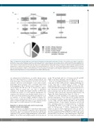
Figure 1. Comprehensive genetic diagnosis of acute myeloid leukemia by next-generation sequencing. (A) Outline of the workflow: each sample is subjected to preparation of three sequencing libraries. Libraries are indexed separately for sequencing on the same flowcell. Data are analyzed using five distinct algorithms for the detection of CNV, fusions, and DNA variants. The whole workflow can be completed within 5 days if performed by one person; times to perform individual steps of the composite assay are indicated on the right. (B) Outline of the CAI[N] algorithm for CNV analysis. Reads are mapped to 1 Mb fixed genomic windows and read distributions are compared to the average of more than 2,500 normal karyotypes (Nfemale=2,819, Nmale=2,605) generated by random sampling of 150-250 bp reads from the reference genome. A region is called amplified or deleted if the observed read number in a window differs significantly (P<0.003) from the average of in silico-generated karyotypes. (C) Flow cell occupancy by three sequencing libraries. Two samples can be analyzed in parallel in one sequencing run on a standard MiSeq v2 flowcell when libraries are sequenced with the read numbers indicated in (A).
we analyzed read distributions on whole chromosomes and in 1 Mb windows for random normal female and male karyotypes. Read frequencies showed very narrow variances and more reads mapped to autosomes in male karyotypes than in female ones, consistent with fewer reads mapping to the Y chromosome compared to a sec- ond X (Figure 2B). Of note, the Y chromosome appears smaller than its actual size, also due to repetitive sequences. To further investigate whether lc-WGS data resemble the results of in silico random experiments, we sequenced two libraries from healthy female donors at 1- 4 x10⁶ reads. Read distribution patterns matched the in sil- ico reference at all read depths examined (Figure 2C). These results confirm that lc-WGS can be accurately sim- ulated computationally, allowing us to use random normal karyotypes as a stable reference for CNV analyses.
Detection of chromosomal gains and losses by copy number variation karyotyping
After evaluation of CAI[N] for consistency with normal karyotypes, we determined its capacity to detect numerical aberrations. First, we examined an individual with Down syndrome (T21) and the benign meningioma cell line BEN- MEN-118 by lc-WGS and CAI[N] analysis. Both trisomy 21
in the T21 proband and loss of chromosome 22 in BEN- MEN-1 cells were identified correctly (Figure 3).
Next, we investigated deletions or additions of chromo- some parts in three AML patients’ samples exhibiting loss of the long arm of chromosome 5 (Table 1, Online Supplementary Figures S1-S3). CAI[N] recovered 5q dele- tions with different breakpoints that closely matched refer- ence laboratory results (Figure 4A, Table 1). Moreover, a gain of chromosome 1p was detected in patient AML-1, consistent with a previously reported partial trisomy 1p (Figure 4B, Online Supplementary Figure S1, Table 1).
Finally, to test the capability of our approach to identify chromosomal gains or losses that are not readily detected by cytogenetic banding, we performed CNV karyotyping on two AML cell lines, HL-60 and NB-4. We observed complex patterns of copy number alterations in both cell lines, including massive overrepresentation of 8q24.21 (containing the MYC locus) with loss of the remaining parts of chromosome 8 (Online Supplementary Figures S4A,B, S5 and S6, Online Supplementary Tables S5 and S6), as described previously.19–26 Amplification of MYC was val- idated by quantitative PCR in HL-60 and NB-4 cells and in patient AML-2, in whom a copy number gain of 8q24 had not been observed by cytogenetics. Similarly, quantitative
haematologica | 2019; 104(2)
279
