
Page 47 - 2019_02-Haematologica-web
P. 47
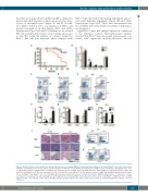
Rheb1 regulates HSC proliferation & differentiation
disorders were apparent in both BM and PB as defined by myeloid left shift (presence of blasts, promyelocytes, mye- locytes, or metamyelocytes) (Figure 4C and D). In addi- tion, Rheb1 deletion led to an expansion of HSCs and myeloid progenitors in BM (Figure 4E-G and Online Supplementary Figure S4C and D). Furthermore, we isolated BM cells and performed serial colony forming unit assays in vitro. We found the number of colonies formed by Rheb1Δ/Δ BM cells was increased when compared with
Figure 4. Rheb1 deletion leads to progressive myeloproliferation in bone marrow (BM) and extramedullary hematopoiesis in aged Rheb1Δ/Δ mice. (A) Survival curves of Rheb1fl/fl and Rheb1Δ/Δ mice (n=22). (B) The absolute number of different cell populations in peripheral blood (PB) of 2-year old Rheb1fl/fl and Rheb1Δ/Δ mice; n=3. (C and D) FACS analysis of granulocytes according to the expression levels of CD11b and Ly-6G in PB and bone marrow (BM); n=3. (E) FACS analysis of LKS+ cells and LKS– in the BM; n=3. (F) The absolute number of LKS+ cells and LKS– in the BM; n=3. (G) The absolute number of GMP, CMP and MEP populations in the BM of 2-year old Rheb1fl/fl and Rheb1Δ/Δ mice; n=3. (H) Whole BM cells were isolated from 2-year old mice and plated in M3434 methylcellulose. Colonies were counted seven days after plating, and serially replating; n=3. (I) Hematoxylin & eosin-stained BM, spleen and liver sections of 2-year old Rheb1fl/fl and Rheb1Δ/Δ mice. (J) FACS analysis of LKS+ cells and LKS– in the spleen. (K) FACS analysis of granulocytes in the spleen. Data are presented as mean±Standard Error of Mean. *P<0.05; **P<0.01.
Rheb1fl/fl BM cells both in first plating experiment and sec- ond serial replating experiment (Figure 4H and Online Supplementary Figure S4E). These data demonstrated that loss of Rheb1 increased proliferation ability of hematopoi- etic progenitor.
Aged Rheb1Δ/Δ mice had enlarged spleens in comparison to the littermate controls. Histopathological analysis revealed that Rheb1Δ/Δ mice developed splenomegaly, con- sistent with significant myeloproliferation disorder
AB
CDE
FGH
IJ
K
haematologica | 2019; 104(2)
251
