
Page 48 - 2019_02-Haematologica-web
P. 48
X. Wang et al.
AB
C
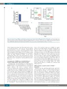
Figure 5. The low expression of RHEB is correlated with poor survival of acute myeloid leukemia (AML) patients with normal karyotype. (A) The percentage of loss- of-function mutations of RHEB in AML patients (http://www.cbioportal.org/). (B) The expression of RHEB in two sets of high-risk versus low-risk AML patients (http://bioinformatica.mty.itesm.mx/SurvExpress; GSE5122 and GSE12417-GPL97). (C) Cumulative (Cum) overall survival of AML patients with different expression levels of RHEB (http://www.abren.net/PrognoScan-cgi/; GSE12417). Data are presented as mean±Standard Error of Mean. ***P<0.001.
(Online Supplementary Figure S4F). Histopathological analy- sis revealed that Rheb1Δ/Δ mice had not only prominent myeloproliferative features including extramedullary hematopoiesis in the spleen, but also infiltration of liver and neutrophilia in BM (Figure 4I). FACS analysis of the spleen revealed a significant enlargement of both the HSC (LKS+) and HPC populations (Figure 4J and Online Supplementary Figure S4G), and with granulocytic expan- sion (Figure 4K). Collectively, these data demonstrate that Rheb1 loss leads to progressive myeloproliferation disor- der in vivo.
Low expression of RHEB was correlated with poor survival in AML patients with normal karyotype
The phenotypes observed in Rheb1 loss and progressive myeloproliferative disorder prompted us to investigate if RHEB also plays a role in human leukemia or myeloprolif- erative diseases. We used a curated database (http://www.cbioportal.org/), which provides large-scale can- cer genomics data sets, to analyze the mutations and copy number alteration (CNA) of RHEB in leukemia patients. It was found that deep deleted mutations in RHEB were about 3% (6 of 188) in an AML patient cohort (Figure 5A). We also analyzed the mutation types in ICGA (interna- tional cancer genome consortium; http://dcc.icgc.org/) and found that the percentage of loss-of-function mutations in RHEB was 1.7% (2 of 117) in AML patients and 0.92% (2 of 218) in CLL patients (Online Supplementary Figure S5A). We then compared the expression of RHEB in AML patients with high-risk versus AML patients with low risk in SurvExpress (http://bioinformatica.mty.itesm.mx/Surv
Express). We found the expression of RHEB was signifi- cantly lower in AML patients with high risk than that of the low risk in two GSE set analyses (GSE5122 and GSE12417-GPL97; P<0.0001) (Figure 5B). We then collect- ed prognostic values for 163 AML patients with normal karyotype in PrognoScan database (http://www.abren.net/ PrognoScan-cgi/, GSE12417), and divided these patients into two groups for analysis: patients with RHEB expres- sion above median and patients with RHEB expression below median (Online Supplementary Figure S5B). The sur- vival curve showed that lower RHEB expression was asso- ciated with poor survival in AML patients with normal karyotype (log-rank test; P=0.034) (Figure 5C). These results indicated that loss of RHEB was correlated with AML progression.
Δ/Δ
Intrigued by the underlying phenotypic plasticities observed in Rheb1Δ/Δ HSCs, we further explored the poten- tial transcriptional changes associated with the pheno- types by microarray analysis. Differentially expressed genes between Rheb1Δ/Δ and Rheb1fl/fl HSCs were signifi- cantly enriched in pathways involved in cell adhesion and cell development (Figure 6A, and Online Supplementary Tables S1 and S2), suggesting that complete loss of Rheb1 in hematopoietic stem cells affected the expression of genes involved in HSCs engraftment and differentiation. Gene set enrichment analysis (GSEA) of pairwise compar- isons revealed that Rheb1Δ/Δ HSCs had a significant increase in the expression of genes associated with AML incidence
3BDO partially rescued the defect of Rheb1 neutrophils
252
haematologica | 2019; 104(2)
