
Page 46 - 2019_02-Haematologica-web
P. 46
X. Wang et al.
A BCD
EFG H
IJKL
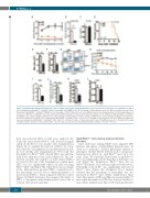
Figure 3. Rheb1 deletion impaired hematopoietic stem cell (HSC) regeneration upon transplantation. (A) Donor-derived-cell chimerism in peripheral blood (PB) of mice transplanted with Rheb1fl/fl or Rheb1Δ/Δ whole bone marrow cells (WBMC); n=6. (B) Percentage of donor-derived-cells in the BM four months after mice were transplanted with Rheb1fl/fl or Rheb1Δ/Δ WBMCs; n=6. (C) Homing of CFSE+ 7-AAD- LKS+ cells to the BM 24 hours (h) post transplantation; n=3. (D) Kaplan-Meier sur- vival curve of Rheb1fl/fl and Rheb1Δ/Δ mice that underwent sublethal irradiation; n=12. (E and F) Absolute number of cells (CD45.1+) in BM and thymus of Rheb1fl/fl and Rheb1Δ/Δ mice under sublethal irradiation; n=3. (G) Percentage of B cells (B220+ ) from the spleens and BM of mice under sublethal irradiation; n=3. (H) Donor- derived-cell chimerism in the PB of mice transplanted with Rheb1fl/fl or Rheb1Δ/Δ LKS+ cells; n=6. (I) Percentage of donor-derived cells in the BM four months after mice were transplanted with Rheb1fl/fl and Rheb1Δ/Δ LKS+ cells; n=6. (J) Absolute number of donor-derived LKS+ cells in the BM per mouse; n=6. (K and L) Self-renewal and differentiation quotients of donor-derived LKS+ cells; n=6. Data are presented as mean±Standard Error of Mean. *P<0.05; **P<0.01;
their donor-derived HSCs in BM were analyzed. We found the donor-derived Rheb1Δ/Δ cells decreased signifi- cantly in the BM at four months after transplantation (Figure 3I). To quantify the function of HSCs, we calcu- lated the LKS+ cell amplification and differentiation abil- ity four months after transplantation.20 We found that the number of Rheb1Δ/Δ LKS+-derived HSCs was substan- tially lower than that of the control (Figure 3J). The self- renewal quotient (the number of donor-derived HSCs recovered at the end of the transplant per original input HSC) was also significantly reduced in mice receiving Rheb1Δ/Δ LKS+ cells (Figure 3K), while there was no change in the differentiation quotient (WBC count/mL blood x the percentage test-cell blood chimerism/number of donor-derived HSCs) when compared with the control (Figure 3L). Rheb1 deficiency thus impaired the ability of HSCs to repopulate under hematopoietic stress due to reduced self-renewal capability.
Aged Rheb1Δ/Δ mice develop myeloproliferative disorders
Since adult mice lacking Rheb1 have impaired HSC function and splenic extramedullary hematopoiesis, we went on to perform a detailed phenotypic analysis to investigate whether Rheb1 deletion leads to progressive hematopoiesis defects in aged Rheb1Δ/Δ mice up to two years of age. We found the survival time of aged Rheb1Δ/Δ mice was significantly shorter than that of littermate con- trols (Figure 4A). Furthermore, Rheb1Δ/Δ mice, but not Rheb1fl/fl littermates, had evident progressive leukocytosis. Neutrophil count in PB was significantly increased in Rheb1Δ/Δ mice (Figure 4B). Peripheral blood analysis revealed that the percentage of neutrophils was also increased in Rheb1Δ/Δ mice (Online Supplementary Figure S4A). The number of RBC and Hgb was normal in Rheb1Δ/Δ mice while the number of PLTs was decreased in Rheb1Δ/Δ mice (Online Supplementary Figure S3B). Myeloproliferative
250
haematologica | 2019; 104(2)
