
Page 45 - 2019_02-Haematologica-web
P. 45
Rheb1 regulates HSC proliferation & differentiation
ABC
DEF
GH
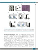
Figure 2. Rheb1Δ/Δ mice display extramedullary hematopoiesis in the spleen. (A and B) Size and weight of spleens in 8-week old Rheb1fl/fl and Rheb1Δ/Δ mice. Data are presented as mean±Standard Error of Mean (SEM); n>6. (C) Hematoxylin & eosin-stained spleen sections of Rheb1fl/fl and Rheb1Δ/Δ mice. Arrowheads indicate
megakaryocytes; arrows point to hematopoietic islands. (D-F) Percentages of LKS+ subsets, LKS– subsets and corresponding subpopulations in Rheb1fl/fl and Rheb1Δ/Δ spleens; n=3. (G) Percentages of B cells (B220+), T cells (CD3+) and myeloid cells (CD11b+) in Rheb1fl/fl and Rheb1Δ/Δ spleens; n=3. (H) Percentages of neutrophils in Rheb1fl/fl and Rheb1Δ/Δ spleens; n=3. Data are presented as mean±SEM. *P<0.05; **P<0.01; ***P<0.001.
Rheb1 deletion impaired HSC regeneration ability in transplant assay
pared with Rheb1fl/fl HS/PCs at 21 days after 4 Gy irradia- tion (Online Supplementary Figure S3B). Thus, a delicate balance of developmental decisions for HSC homeosta- sis, including stem cell quiescence, self-renewal and dif- ferentiation, was maintained under steady state condi- tions in Rheb1Δ/Δ mice but was broken under hematopoi- etic stress.
We further examined the self-renewal and differentia- tion capacities of HSCs using LKS+ transplantation. A total of 200 LKS+ cells isolated from Rheb1fl/fl and Rheb1Δ/Δ mice (CD45.1) together with 5x10⁵ WBMCs (CD45.2) were intravenously injected into lethally irradiated recip- ient mice. The chimerism in PB was analyzed every four weeks post transplantation. The repopulating capacity of Rheb1Δ/Δ LKS+ cells in the PB was significantly lower in recipient mice than the control LKS+ cells (Figure 3H). All lineages derived from Rheb1Δ/Δ HSCs were significantly reduced in the PB and the BM after transplantation (Online Supplementary Figure S3C). The recipient mice were sacrificed at four months after transplantation and
To assess the role of Rheb1 in HSC function during hematopoiesis, we transplanted Rheb1Δ/Δ BM cells (CD45.1) with competitive cells (CD45.2) into lethally irradiated recipient mice (CD45.2) to examine the role of Rheb1 in adult HSC reconstitution. All lineages derived from Rheb1Δ/Δ cells were significantly reduced in the PB and the BM after transplantation (Figure 3A and B, and Online Supplementary Figure S3A), while the homing capacity of transplanted Rheb1Δ/Δ CFSE+ labeled cells was equivalent to that of Rheb1fl/fl cells (Figure 3C).
In addition, Rheb1fl/fl and Rheb1Δ/Δ mice were subjected to 4 Gy of X-ray irradiation and we examined the effects of Rheb1 deficiency on HSCs under hematopoietic stress. Interestingly, all Rheb1fl/fl mice survived while most Rheb1Δ/Δ mice died, probably due to impaired recovery of the BM and thymic cellularity (Figure 3D-F) and impaired B lymphopoiesis in the BM and spleen (Figure 3G). The absolute number of Rheb1Δ/Δ HS/PCs was reduced com-
haematologica | 2019; 104(2)
249
